Gastric Volvulus
Michael P. Federle, MD, FACR
Key Facts
Imaging
Organoaxial volvulus: Rotation of stomach around its longitudinal axis
Most common type
Stomach twists either anteriorly or posteriorly
Antrum moves from inferior to superior
Mesenteroaxial volvulus: Rotation of stomach about mesenteric axis
Stomach rotates from right to left or left to right about long axis of gastrohepatic omentum
Entire stomach may be herniated (type 4 paraesophageal hernia) or only part (type 3 PEH)
Either can result in volvulus, ± obstruction, ± ischemia
Gastric wall pneumatosis = ischemia
CT chest and abdomen; performed preoperatively
To detect associated malformation or malposition and site, size, level of diaphragmatic defect
Top Differential Diagnoses
Hiatal hernia
Type 3 & 4 paraesophageal hernias at risk for GV
Postoperative state, stomach
Esophagectomy with gastric pull through (conduit may twist and obstruct)
Pathology
Associated abnormalities
Large paraesophageal hernia
Diaphragmatic eventration or paralysis
Hernia of colonic transverse loop ± other bowel loops
Diagnostic Checklist
Presence or absence of obstruction and ischemia are more important than remembering or reporting whether volvulus is organo- or mesenteroaxial
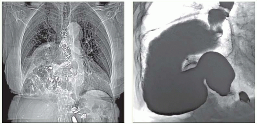 (Left) Chest film in an elderly woman shows stomach and other bowel within the right hemithorax. (Right) An upper GI series in the same patient confirms an organoaxial volvulus of the stomach. This is a type 4 paraesophageal hernia (intrathoracic stomach). There is minimal obstruction at the time of this study, but the patient is at risk for strangulation and obstruction. |
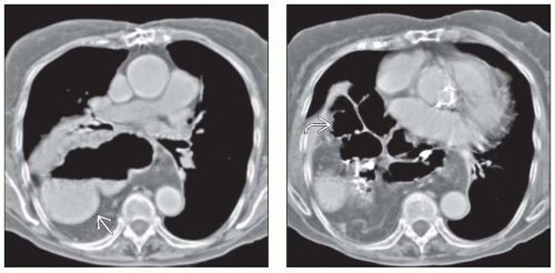 (Left) CT in the same patient shows the stomach  within a hernia sac in the right thorax, and it is rotated on its long axis (organoaxial volvulus). (Right) Much of the transverse colon within a hernia sac in the right thorax, and it is rotated on its long axis (organoaxial volvulus). (Right) Much of the transverse colon  is also within the hernia sac in the same patient. Due to the enormous size of the hiatal defect in the diaphragm, neither the stomach nor the colon was pinched as it traversed the diaphragm. is also within the hernia sac in the same patient. Due to the enormous size of the hiatal defect in the diaphragm, neither the stomach nor the colon was pinched as it traversed the diaphragm. |
TERMINOLOGY
Abbreviations
Gastric volvulus (GV)
Definitions
Uncommon acquired twist of stomach on itself
IMAGING
General Features
Morphology
Abnormal degree of rotation of 1 part of stomach around another part
Types of GV: Organoaxial (most common), mesenteroaxial, mixed
Organoaxial volvulus (OAV): Rotation of stomach around its longitudinal axis
Around line extending from cardia to pylorus
Stomach twists either anteriorly or posteriorly
Antrum moves from inferior to superior position
Mesenteroaxial volvulus (MAV): Rotation of stomach about mesenteric axis
Axis running transversely across stomach at right angles to lesser and greater curvatures
Stomach rotates from right to left or left to right about long axis of gastrohepatic omentum
Mixed volvulus: Combination of OAV & MAV
Radiographic Findings
Radiography
Abdominal plain films; patient upright
Double air-fluid level
Large, distended stomach; seen as air- and fluid-filled spheric viscus displaced upward and to left
Small bowel collapsed if stomach is obstructed
Chest film: Intrathoracic; upside-down stomach
Retrocardiac fluid level; 2 air-fluid interfaces at different heights; suggests intrathoracic GV
Fluoroscopic Findings
Massively distended stomach in left upper quadrant extending into chest
Inversion of stomach
Greater curvature above level of lesser curvature
Positioning of cardia and pylorus at same level
Downward pointing of pylorus and duodenum
OAV: 2 points of twist; luminal obstruction
Incomplete or absent entrance of contrast material into &/or out of stomach; acute obstructive GV
OAV: Failure of contrast to enter stomach; obstruction at esophagus or proximal stomach
If contrast material does enter stomach, it may not pass beyond obstructed pylorus
May see “beaking” at point of twist
MAV: Antrum and pylorus lie above gastric fundus
CT Findings
CT appearance may be variableRelated posts:

Stay updated, free articles. Join our Telegram channel

Full access? Get Clinical Tree


