Chapter 3
Gastrointestinal, Gynaecological, and Urological Anatomy
 Question 1:
| Name the arrowed structure |
 Question 2:
| Name the arrowed structure |
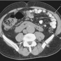
 Question 1: Axial CT of the abdomen
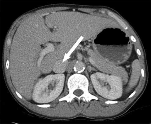
Answer: Inferior vena cava
• The inferior vena cava (IVC) passes adjacent to to the posterior part of the liver.
• The IVC has a short intrathoracic course. This may be important when it comes to answering the question in the examination, ‘intrathoracic inferior vena cava’, if the marker points to the IVC above the diaphragm.
• In the image, the IVC is distended and appears larger than the aorta. Sometimes it can appear slitlike when it is collapsed; it varies with the phase of respiration and intravascular volume. Be sure to familiarise yourself with both appearances.
• A bilateral/double IVC is a well-described normal variant.
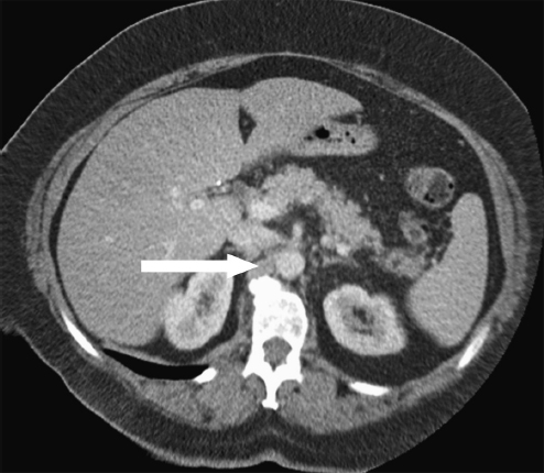
 Question 2: Axial CT of the abdomen
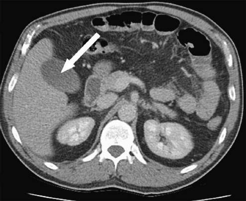
Answer: Gallbladder
• The gallbladder is a fluid-filled structure and therefore has a lower attenuation than the adjacent liver.
• Rarely, it may be intrahepatic, surrounded by liver parenchyma rather than on the undersurface of the liver.
• Its wall should be less than 3 mm in thickness.
• Gas within the gallbladder may be a normal finding shortly after sphincterotomy or biliary stent insertion.
 Question 3:
| Name the arrowed structure |
 Question 4:
| Name the arrowed structure |
 Question 3: US of the abdomen
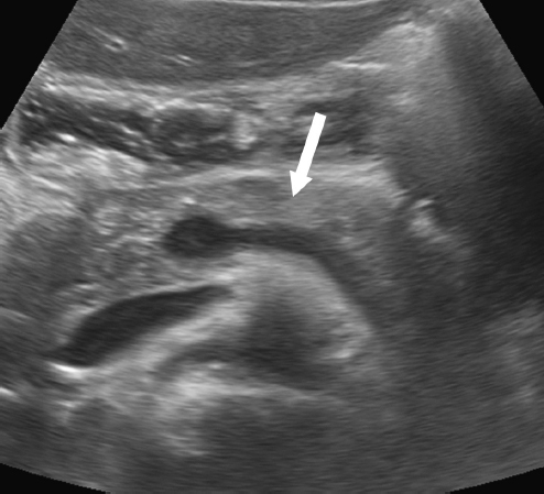
Answer: Body of the pancreas
• The pancreas is divided into an uncinate process, head, neck, body, and tail.
• Only the neck has an anatomical definition; it is located anterior to the confluence of the superior mesenteric vein and splenic vein.
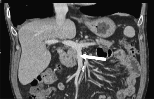
 Question 4: Coronal CT of the abdomen
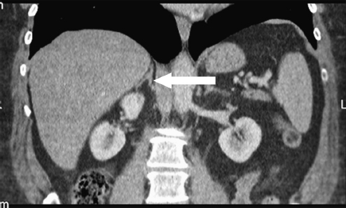
Answer: Medial limb of the right adrenal gland
• The right adrenal gland is usually found cranial to the right kidney, whereas the left adrenal gland lies anteromedial to the upper pole of the left kidney.
• The normal thickness of a limb of the adrenal gland is approximately the same thickness as the adjacent diaphragm.
• The adrenal gland is composed of the body and the medial and lateral limbs. If in doubt as to how specific you should be in the examination, you should provide more information and specify the part of the adrenal gland that is labeled.
 Question 5:
| Name the arrowed structure |
 Question 6:
| Name the arrowed structure |
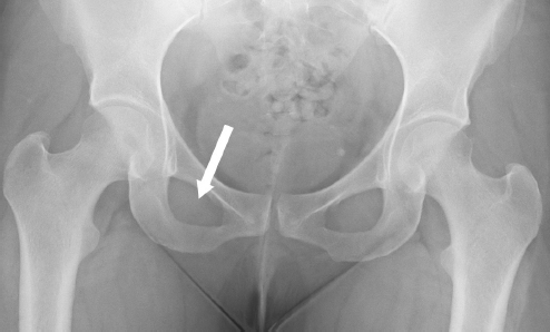
 Question 5: AP radiograph of the pelvis
Answer: Right obturator foramen
• The obturator foramen is bordered by the pubic rami and the ischial bone.
• The foramina are almost entirely covered by a ligamentous membrane.
• The obturator externus muscle originates from the external surface of the obturator membrane. The obturator internus muscle originates from the deep surface and hooks around the pelvic bones to insert onto the femur.
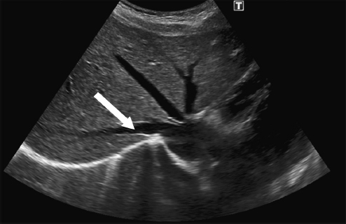
 Question 6: Transverse US of the abdomen
Answer: Right hepatic vein
• The venous drainage of the liver is variable, but typically the majority of the liver is drained by three hepatic veins draining into the intrahepatic portion of the inferior vena cava (IVC).
• The caudate lobe drains directly into the IVC, which explains why this part of the liver is often not affected by disease processes in the same way as the remainder of the liver.
• The right hepatic vein is one of the landmarks used to separate segments V and VI as well as VII and VIII of the right lobe of the liver.
 Question 7:
| Name the arrowed structure |
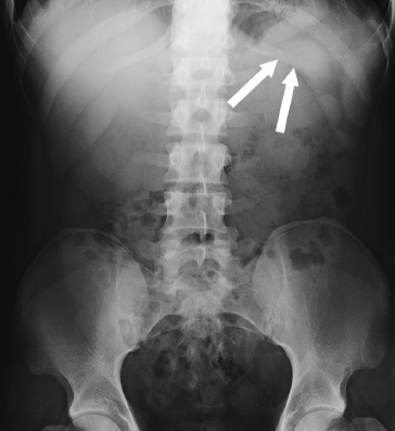
 Question 7: AP radiograph of the abdomen
Answer: Left 12th rib
• The 12th rib is one of the free floating ribs, which means that it is not attached anteriorly to the sternum.
• It can be seen on abdominal radiographs as the most inferior rib, tapering after a few centimetres.
• The 12th ribs can be helpful when counting the vertebral levels of the lumbar spine because their origin defines the T12 vertebral level.
• The 12th ribs can vary in length and may be absent.
 Question 8:
| Name the arrowed structure |
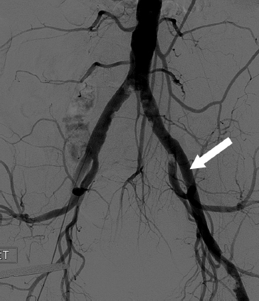
 Question 8: Angiogram of the iliac arteries
Answer: Left external iliac artery
• The common iliac arteries divide into the internal and external iliac arteries.
• The external iliac arteries run parallel and usually lateral to the external iliac veins.
• The external iliac artery becomes the femoral artery as it passes the inguinal ligament. This is a common pitfall in the examination.
 Question 9:
| Name the arrowed structure |
 Question 10:
| Name the arrowed structure |
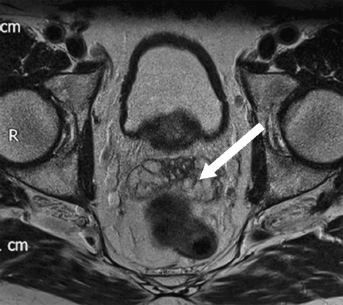
 Question 9: Axial T2-weighted MRI of the male pelvis
Answer: Left seminal vesicle
• The seminal vesicles are paired structures posterior to the male bladder.
• Due to the high water content of the vesicles, they are of high signal intensity on T2-weighted MRIs.
• It is possible to confuse the seminal vesicles with ovaries when looking at just one slice. The way to determine whether this is a male or female pelvis is to look for the presence of the uterus—not here in this example—and to look for the presence of the prostate gland, situated behind the bladder as in this image.
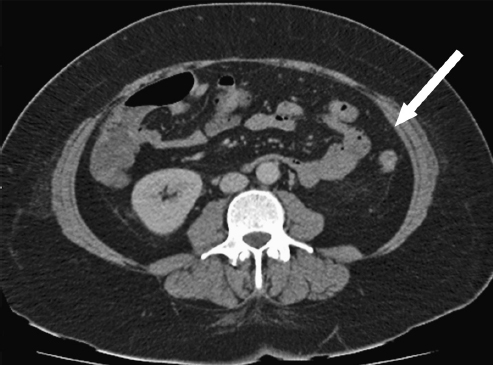
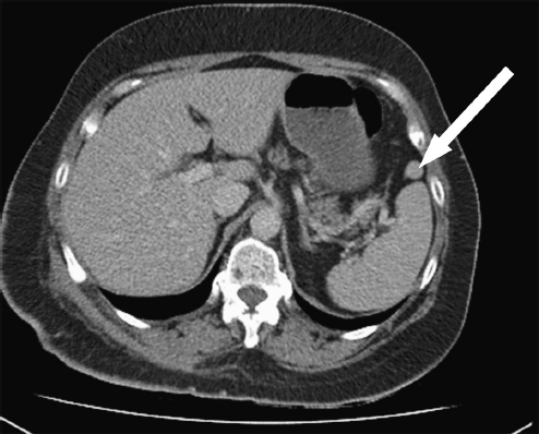
 Question 10: Axial CT of the abdomen
Answer: Left transversus abdominis muscle
• The left transversus abdominis muscle is the innermost of the lateral abdominal muscles. Its fibres run transversely from lateral to medial.
• On its inferior border, it forms a tendinous fold, part of which is the roof of the inguinal canal.
 Question 11:
| Name the arrowed structure |
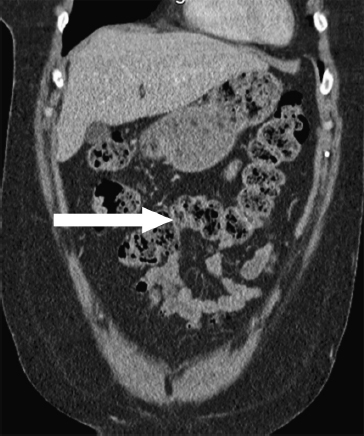
 Question 11: Coronal CT of the abdomen
Answer: Transverse colon
• The transverse colon is the most distal part of the colon to be supplied by branches from the superior mesenteric artery.
• It is suspended from the transverse mesocolon and lies within the peritoneal cavity.
• It is of variable length and may be seen drooping as far down as the pelvis.
 Question 12:
| Name the arrowed structure |
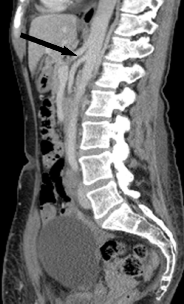
 Question 12: Sagittal CT of the abdomen and pelvis
Answer: Coeliac trunk
• The coeliac trunk is the first anterior branch of the abdominal aorta and supplies the foregut.
• It gives rise to the common hepatic, splenic, and left gastric arteries.
 Question 13:
| Name the arrowed structure |
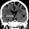
 Question 13: Axial CT of the abdomen
Answer: Right crus of the diaphragm
• The diaphragmatic crura are a paired structure; however, the right crus extends more inferiorly to L3 and may be confused with other structures.
• The left crus inserts onto L2.
 Question 14:
| Name the arrowed structure |
 Question 15:
| Name the arrowed structure |
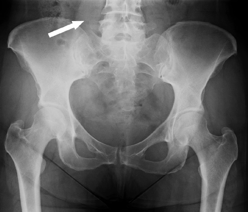
 Question 14: AP radiograph of the pelvis
Answer: Right psoas major muscle
• The psoas major muscle may be seen on plain radiographs as an elongated, wedge-shaped density running obliquely next to the lumbar spine.
• It is one of the flexors of the hip, originating from the lateral border of the spine (T12 to L5) and inserting into the lesser trochanter of the femur.
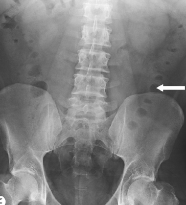
 Question 15: AP radiograph of the abdomen
Answer: Descending colon
• The descending colon is often seen as a gas-filled structure on plain radiography, running in the superoinferior direction in the left flank.
• It can be identified as large bowel by its haustral pattern and faeces may be seen within.
• The upper limit of normal for the descending colon is 6 cm in diameter. The caecum may measure up to 9 cm and small bowel up to 3 cm.
 Question 16:
| Name the arrowed structure |
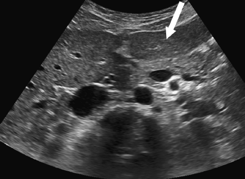
 Question 16: Transverse US of the upper abdomen
Answer: Lateral segment of the left lobe of liver
• The functional left lobe of the liver lies to the left of the Cantlie line. This is the plane between the inferior vena cava (IVC) and the gallbladder fossa. The left lobe consists of segments I to IV—named by Couinaud.
• It crosses the midline and lies under the diaphragm with the heart superiorly, the stomach and duodenum inferiorly, and aorta and IVC posteriorly.
• The left lobe is supplied by the left portal vein and left hepatic artery. Venous drainage is by the left hepatic vein, and biliary drainage is via the left hepatic duct.
• At first glance, the image is disorientating because only a part of the left and right lobes of the liver can be seen. However, if you look at the rest of the image, you will notice the IVC and aorta are sitting anterior to the vertebral body in the bottom central part of the image. You will also notice the fissure for the falciform ligament, which divides the left lobe into medial and lateral segments. More deeply is the pancreas, which is more reflective than the liver, and the splenic vein posteriorly.
• The figure below shows the segments of the liver as described by Couinaud.
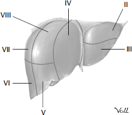
From Atlas of Anatomy, © Thieme 2008, illustration by Markus Voll.
 Question 17:
| Name the arrowed structure |
 Question 18:
| Name the arrowed structure |
 Question 17: Coronal CT of the abdomen
Answer: Superior mesenteric vein
• The superior mesenteric vein (SMV) is the principal venous drainage of the bowel. It joins the splenic vein to form the portal vein.
• The SMV runs to the right of the superior mesenteric artery (SMA). Reversal of the SMA/SMV relationship is a feature of intestinal malrotation.
• The confluence of the SMV and splenic vein defines the neck of the pancreas.
Related posts:
Stay updated, free articles. Join our Telegram channel

Full access? Get Clinical Tree


