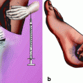
Fig. 2.1
The ultrasound scan of fetal head During the labor, in longitudinal and transverse section and in soprapubic (or transabdominal) subpubis (or translabial) scan. (a) the figures shows the soprapubic sagittal scan of the fetal head. (b) the figures shows a transverse scan of fetal head sovprabubic. (c) The figures show the subpubic sagittal (or translabial) scan of fetal head. (d) the figures show the transversal sonogram of fetal head (or translabial) under the simphisis
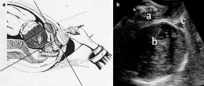
Fig. 2.2
(a) Sagittal section of female pelvis at term of pregnancy in the first stage of labor and transvaginal sonography: the lines passing through the scan represent the main longitudinal and transverse scans. (b) Image obtained after intrapartum sagittal medial translabial exploration; “a” indicates the maternal pubis, “b” indicates the fetal head, and “c” indicates the fetal caput succedaneum
Ghi et al. [4] suggest a series of settings for the acquisition of adequate intrapartum fetal head and maternal pelvis volumes (Table 2.1).
1 – Lowest possible angle of insonation |
2 – Lower output frequency |
3 – Highest insonation depth |
4 – Wide volumetric area with low sound volume |
2.2.5 Determining the Type of Exploration
Ultrasound exploration in the delivery room can be useful for two purposes: first, to document the fetal head position in relation to the maternal pelvis (e.g., before an instrumental delivery), and second, to determine labor progression. These two purposes are undoubtedly the parameters of most interest in the intrapartum study. Fetal head descent in the maternal pelvis, rotation, and fetal head direction can be evaluated to determine whether labor is progressing adequately.
2.3 Ultrasound Use in Labor
The use of intrapartum ultrasound has been widely discussed in the last five years. Different studies discuss the uses of ultrasound in the delivery room [1, 5]. Some of these uses are considered basic and do not require a significant level of specialization in obstetric sonography [1], such as with excluding or confirming fetal death, determining fetal status, determining fetal biometry to estimate approximate weight, and determining the degree of flexion of the fetal head. When describing the uses of ultrasound in the delivery room, these indications cannot be forgotten; however, they will not be the most important indications when a patient is in labor [1]. Other uses of intrapartum sonography, such as amniotic fluid evaluation and placenta localization, should be conducted within fetal pathophysiology and not within the delivery room [1]. In Table 2.2, we provide a classification system that we created to classify the uses of intrapartum sonography [1]. Practically speaking and to the best of our knowledge, ultrasound use during labor can be summarized as serving two purposes: determining fetal head position in relation to the maternal pelvis and objectively documenting inadequate progression of labor.
1. Basic uses |
Identification of fetal heartbeat |
Determination of fetal presentation |
Attainment of somatometry; determination of estimated fetal weight (to detect fetal macrosomia) |
Placental location |
Determination of the degree of cephalic flexion (podalic birth) |
Assistance in the birth of the second twin |
Postpartum metrorrhagia |
2. Advanced uses |
Determination of fetal head position |
Diagnosis of stalled labor |
Assessment of intrapartum fetal well-being (intrapartum Doppler) |
2.3.1 Ultrasound Use to Determine Fetal Head Position
The digital determination of the fetal head position using an obstetric exploration that requires training is greatly influenced by the laboring woman’s degree of cervical dilation and has added difficulties with the presence of caput succedaneum or asynclitism. Some studies have shown that the digital determination of fetal head position with respect to the maternal pelvis during labor is not exact [6, 7]. Likewise, Akmal et al. [8] conducted a study of 496 abdominal ultrasounds after digital exploration and found that fetal head position could not be determined via vaginal touch in 33% of the cases, especially in cases with occipitoposterior positions [9]. Souka et al. [10] could not determine fetal head position in 61% of subjects during the first stage of labor and in 31% during the second stage. In a study of 44 pregnant women in the intrapartum setting, Kreise et al. [11] found erroneous interpretations of fetal head position in 30% of cases after digital exploration. Dupuis et al. [12] found a 70% concordance of both methods in determining fetal head position, with more difficulties observed in cases with caput succedaneum and posterior positions.
Chou et al. [13] found greater precision for determining occipitoposterior positions with transabdominal ultrasound than with vaginal touch (92% vs. 72%, respectively). Zahalka et al. [14] compared the use of vaginal touch, transabdominal ultrasound, and transvaginal ultrasound to determine fetal head position and found that transvaginal ultrasound was the most exact method. In our center, which had an average of 7,900 births in the last 10 years and an average C-section rate lower than 12%, we believe that there is no substitute for adequate clinical management; however, the majority of delivering obstetricians find that transabdominal or translabial ultrasound is useful for determining fetal head position, primarily at the point at which an instrumental delivery is indicated [15]. In other centers with perhaps less clinical experience, ultrasound could be useful for decreasing the number of C-sections performed defensively by obstetricians who wish to avoid the possibility of a complicated delivery [16].
In a recently published article, we evaluated the usefulness of transabdominal ultrasound to determine fetal head position during the first and second stages of labor in 86 consecutive patients [1]. With the patient in the decubitus supine position or the dorsal lithotomy position after bladder-emptying, we placed the abdominal probe transversally in the suprapubic area. The fetal head position was defined by the identification of the orbits (occipitoposterior), midline echo (occipitotransverse), and cerebellum or column (occipitoanterior) [17] (Fig. 2.3a, b). Using transvaginal exploration followed by transabdominal ultrasound, we did not find significant differences between these methods’ ability to determine the fetal head position. Clinically, it was possible to determine fetal head position in 93% of the patients in the first stage of labor and in 96% of the patients in the second stage of labor, and there was a concordance with the ultrasound results in 98% of the cases. A body mass index in the top 15% of women was the only significant factor that made transabdominal ultrasound exploration difficult, while the presence of caput succedaneum was important for determining fetal head position [1]. Instrumental delivery with forceps or a vacuum requires awareness of the fetal head position because the incorrect application of these instruments can lead to adverse results. Wong et al. [18] randomized 50 patients with a prolonged second stage of labor prior to vacuum extraction to digital pelvic exploration only or transabdominal ultrasound exploration in conjunction with digital exploration. The authors showed that including ultrasound allowed a more precise application of the vacuum extractor. In our studies, we determined the exact clinical and ultrasound fetal head position prior to the application of forceps in 13 cases. In our center, which has a C-section rate of approximately 12% and a forceps delivery rate of 14%, it is imperative to determine the fetal head position for better labor management. In contrast, Akmal et al. [19] studied 64 labors prior to instrumental delivery and found that digital exploration failed to determine the fetal head position in 26% of the cases. The existence of a higher C-section rate or the use of vacuum extraction as the instrumental technique of choice may be responsible for the inaccuracy of fetal head position determination via transvaginal exploration. The differences in the published data demonstrate the need for a prospective study to more precisely specify the usefulness of ultrasound to determine the fetal head position prior to an instrumental delivery.
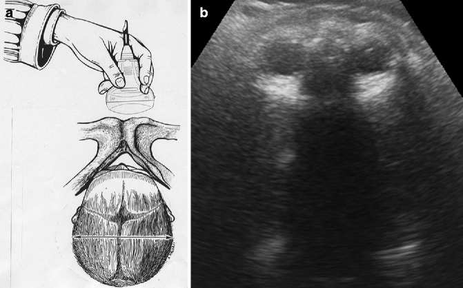
Fig. 2.3
(a) 2D transabdominal ultrasound in cross section during labor with fetal head in median occiput posterior position. (b) Intrapartum transabdominal ultrasound (suprapubic transverse) of a patient in the second stage of labor with the fetus in the occipitoposterior position. Note that both orbits are directed toward the ultrasound transducer
To a lesser extent, studies have also described the determination of fetal head position using 3D transperineal ultrasound [20]. In our center, we obtained 20 captures of sutures and fontanels using translabial ultrasound with a transabdominal probe. Such volumetric captures allow us to identify the sutures and fontanels of the fetal head, which is useful prior to an instrumental delivery or when deciding whether to allow a vaginal delivery attempt or perform C-section (Fig. 2.4a, b).
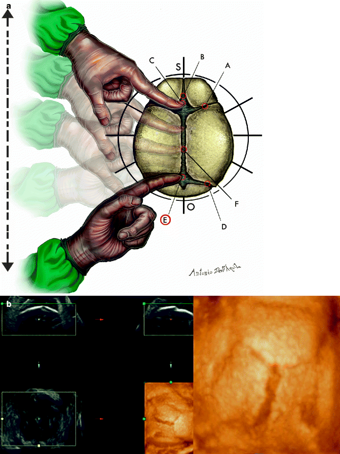
Fig. 2.4
“Drawing of sutures and fontanels and ours digital palpations: (A) coronal sutures; (B) frontal sutures; (C) anterior fontanel (bregma or major fontanel); (D) occipital sutures; (E) “circumference” posterior fontanel or lambdoid suture or small fontanel; (F) parietal suture (or sagittal suture). O occiput, S sinciput. (b) Identification of sutures and fontanels using 3D ultrasound in a patient during the second stage of labor. Note that the lambdoid fontanels are to the right in a fetus with an occipitoanterior position”
The objective information about the fetal head position that 2D or 3D ultrasound provides prior to an instrumental delivery appears to be among the most useful data for decreasing the number of C-sections or adverse results after the inadequate performance of instrumental deliveries.
2.3.2 Ultrasound Use for Diagnosing a Stalled Labor
If the fetal head position is anterior, posterior, or transverse, ultrasound is clearly warranted to determine the exact fetal position before delivery; however, using ultrasound during delivery can also provide objective data for diagnosing inadequate progression of labor [8, 21]. Currently, the number of C-sections is increasing in an unjustified manner, often due to the practice of defensive medicine. We commented previously on the impreciseness of digital touch for the evaluation of the fetal head position relative to the maternal pelvis [22]; the combination of ultrasound exploration with clinical exploration could be useful for decreasing the number of C-sections.
The inaccuracy of clinical examination during labor has also been demonstrated regarding the descent of the head across the different planes of the maternal pelvis. Dupuis et al. [23] investigated the reliability of transvaginal assessment of fetal head station using a newly designed birth simulator. A fetal head mannequin was placed in one of the 11 American College of Obstetricians and Gynecologists (ACOG) stations in a birth simulator equipped with a real-time miniaturized sensor. The operators (32 residents and 25 obstetricians) then determined the head position clinically. The ACOG position was incorrectly determined in 50–88% of cases for residents and in 36–80% of cases for obstetricians, depending on the position. When the stations were examined in groups (high, midpelvis, low, and outlet), the mean group error was 30% (95% CI: 25–35%) for residents and 34% (95% CI: 27–41%) for obstetricians. Alarmingly, the group errors show that the misdiagnosis of a station as midpelvic rather than high-pelvic accounted for 88% and 67% of the errors made by residents and obstetricians, respectively. This misdiagnosis can have serious implications for the management of patients in labor.
To determine the progression of labor using either 2D or 3D sonography, the exploration must be performed translabially. The patient must be placed in a semicurved position with flexed legs, and amniotomy is recommended [24]. The ultrasound transducer is placed in the medial sagittal plane, between the labia majora and precisely below the symphysis pubis. Several studies have used ultrasound to provide an objective measure of head progression in labor:
The most important measurement is the angle of progression of the fetal head, described as the angle between a line through the midline of the pubic symphysis and a line from the inferior apex of the symphysis to the leading part of the fetal skull (Fig. 2.5a, b). An angle of progression of 120° or greater is an excellent predictor of a successful vaginal delivery. Kalache et al. [25] evaluated this measurement prospectively in women at term with failure to progress in the second stage of labor. Logistic regression analysis showed a strong relationship between the angle of progression and the need for cesarean delivery. When the angle of progression was 120°, the fitted probability of either an easy and successful vacuum extraction or a spontaneous vaginal delivery was 90%. The same angle was measured by Barbera et al. [2, 26] in 88 term laboring patients. The authors described a good intra- and interobserver variability for measurements that were less than 3°. Their data showed that an angle of at least 120° was always associated with subsequent spontaneous vaginal delivery.
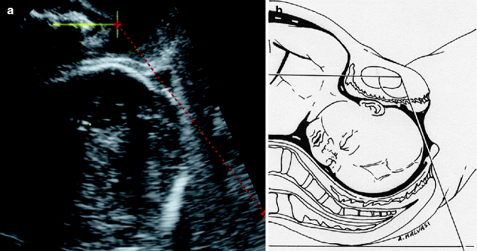
Fig. 2.5
(a) Ultrasound image and drawing demonstrating the angle of fetal head progression, described as the angle between a line through the midline of the pubic symphysis (continuous yellow line) and a line from the inferior apex of the symphysis to the leading part of the fetal skull (interrupted red line). (b) The image shows the equivalent diagram of the angle of the progression of the fetal head
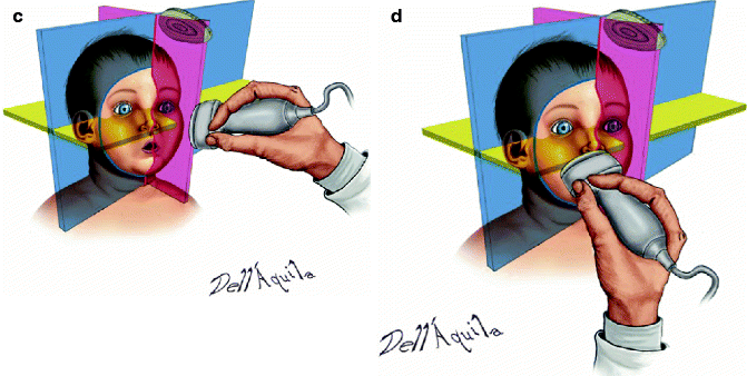
Head direction: Defined by Hernich [27] as the angle between the infrapubic line of the pelvis (a line perpendicular to the longer diameter of the pubis starting from the inferior border) and another line drawn perpendicular to the widest diameter of the fetal head (Fig. 2.6a, b). Using this technique, three types of head directions were determined: head down, horizontal, and head up. “Head up” is when the line perpendicular to the widest diameter of the fetal head points ventrally at an angle of ≥ 30°; “head down” is when this angle is <0°; all other angles are considered horizontal. The head direction, together with the descent in the maternal pelvis, is a good indicator of successful vaginal delivery. An upward direction of the fetal head is a good prognostic sign for vaginal delivery, in contrast with a downward or horizontal head direction [22].
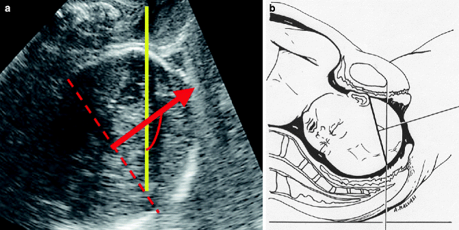
Fig. 2.6
(a) Ultrasound image and drawing demonstrating the fetal head direction, described as the angle between a vertical line from the inferior apex of the symphysis (yellow line) and a line drawn perpendicular to the widest diameter of the fetal head (red arrow). (b) The ultrasound image shows the corresponding angle of the direction of the fetal head in the birth canal
Progression distance: Defined by Dietz et al. [28] as the minimum distance between a line through the inferoposterior margin of the symphysis pubis and the border of the fetal skullcap (Fig. 2.7a, b). This indicator is a useful marker for the determination of fetal head station. Information about the degree of fetal head descent is necessary prior to the use of forceps or a vacuum. It may be helpful to determine these measurements when determining fetal head station [29, 30]. The results of one of our studies comparing all the measurements of 50 women in the second stage of labor indicated that angle progression has the best intra- and interobserver reproducibility when studying fetal head progression during labor [31].Get Clinical Tree app for offline access
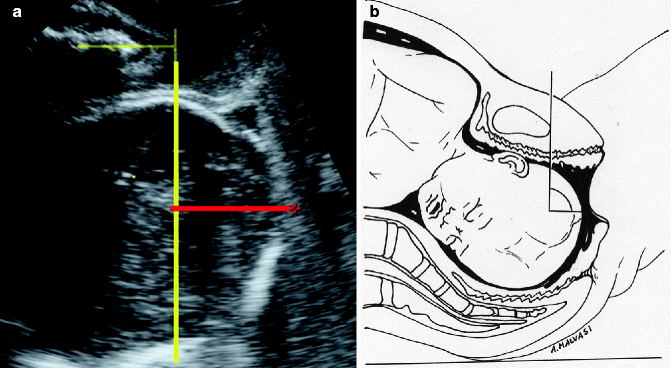
Fig. 2.7