Geriatric Imaging
OBJECTIVES
After studying this chapter, the student will be able to:
1. Discuss the special considerations while imaging geriatric patients and describe their special care needs.
2. Discuss the normal changes associated with aging for the integumentary, cardiovascular, pulmonary, hepatic, gastrointestinal, genitourinary, and musculoskeletal systems.
3. Explain the precautions to be taken for a patient who has had an arthroplasty.
4. Discuss cultural considerations when caring for the geriatric patient.
5. Discuss the radiographer’s role in a suspected elder abuse procedure.
KEY TERMS
Actinic keratosis: A slow, localized thickening of the outer layers of the skin as a result of chronic, excessive exposure to the sun
Adduct: To draw toward a center, or median line
Alzheimer disease: An illness characterized by dementia, confusion, memory failure, disorientation, restlessness, speech disturbances, and an inability to carry out purposeful movements; onset usually occurs in persons aged 55 years or older
Arthroplasty: Plastic repair of a joint
Baroreceptor: A sensory nerve terminal that is stimulated by changes in pressure
Dementia: Organic mental syndrome characterized by general loss of intellectual abilities involving impairment of memory, judgment, and abstract thinking
Depression: A morbid sadness, dejection, or melancholy
Geriatrics: The branch of medicine that deals with all aspects of aging, including pathologic and social problems
Hallucination: A sensory impression (sight, sound, touch, smell, or taste) that has no basis in external stimulation
Kyphosis: An abnormal condition of the vertebral column in which there is increased convexity in the curvature of the thoracic spine
Lentigines: Round, flat, brown, highly pigmented spots on the skin caused by increased deposition of melanin; often occurs on the exposed skin of the elderly
Macule: A small, flat blemish or discoloration that is flush with the skin surface
Polypharmacy: Administration of excessive medications or of many drugs together
Presbyopia: Loss of ability to focus on near objects
Prosthesis: An artificial substitute for a missing part
Urinary incontinence: Inability to control urinary functions
THE GERIATRIC PATIENT
The number of people aged over 65 years in the United States are predicted to be more than 74 million people by the year 2030. At the present time, one in every eight persons falls into this category. With 85 million baby boomers, the largest population reaching 60 years, one of every five Americans is projected to be over 65 by 2030. Persons aged over 85 years constitute one of the fastest-growing portions of the population (Display 8-1). By the year 2030, the population 85 years and over could increase by 21%. For health care workers, this means that most of their patients will be 65 years or older; therefore, they must understand the changes that occur as one ages. The radiographer must understand that age 65 is an arbitrary age that has been designated for convenience as the age at which a person is eligible for Medicare benefits, Social Security benefits, and retirement from many positions. Many persons at this age are still quite youthful and productive; therefore, all persons must be assessed on an individual basis, not merely by their chronologic age (Display 8-2).
DISPLAY 8-1 Aging Americans
| |||||||||||||||||||||
DISPLAY 8-2 Categories of Persons Who Are 65 Years and Older
|
The human body undergoes normal physiologic and anatomic changes as it ages. These changes do not occur uniformly in all people, so it is not correct to say that all persons begin to demonstrate the changes of age at the same time. Lifestyles, culture, and hereditary factors contribute to the aging process. When an elderly patient is admitted to the imaging department, the radiographer must be able to differentiate between the normal changes of aging.
The elderly person is more frequently burdened with major illnesses that are chronic rather than acute in nature. Heart disease, cancer, and strokes are the cause of 80% of deaths in persons aged over 65 years. Hypertension, arthritis, diabetes mellitus, pulmonary disease, and visual and hearing impairments are also common conditions requiring long-term care. These conditions result in a great deal of physical discomfort and a multitude of social and psychological problems.
Large numbers of medications are prescribed and consumed by the elderly. Many of these medications affect the patient’s response level. Often, problems attributed to the aging process are the result of multiple medication consumption, which is frequently called polypharmacy.
Depression is a common and debilitating emotional problem of the aged person. The threat of becoming a burden to one’s family, the fear of losing one’s good health, or the necessity of giving up an independent lifestyle owing to chronic disease result in feelings of helplessness and hopelessness that can give rise to a major depressive episode. Symptoms of depression in the elderly person are often confused with dementia. Symptoms of dementia, including disorientation, confusion, gross memory deficits, paranoid ideation, hallucinations, and depression, are not part of the normal aging process. When these symptoms are present in an elderly patient, the radiographer must remember that they are indicative of a disease process. Persons with Alzheimer disease present with symptoms of dementia, which may occur in persons aged over 65 years, although it may occur at an earlier time. Persons with Alzheimer disease or other diseases that produce symptoms of dementia are frequently seen for a radiographic imaging examination. Recent studies have indicated that of people aged over 85 years about 50% suffer from Alzheimer disease at various stages.
To ensure the correct patient will be imaged, the technologist must implement the two patient identifiers for all patients. When the geriatric patient is confused, it is imperative to verify the patient’s identification with their wrist band, the family member or caregiver, and/or their medical records.
When caring for a patient who exhibits symptoms of dementia, remember that the person may not be able to understand or retain directions. The radiographer will have to explain to the patient what is to be done and then assist him or her in following the directions. The patient who is confused or has a paranoid ideation may become frightened when confronted by the forbidding atmosphere of the radiographic imaging department. Allowing a familiar person to be present in the examination room to assist the patient may make the patient feels more secure and allow the procedure to be accomplished more effectively.
Persons who are depressed may be so preoccupied that they do not respond to outside stimuli. It may be necessary to give the same instructions a number of times to a depressed person and then to assist the patient in following directions.
When an examination of the geriatric patient is complete, the radiographer must assist him or her to return to the dressing room or lavatory, because the stress of the procedure may result in forgetfulness. Make certain that the patient is attended before leaving him or her. The elderly patient must not be left alone in the radiographic imaging room, because he or she may become confused and may be in danger of falling from the examining table.
CHANGES ASSOCIATED WITH AGING
Older adults may not have the same symptoms of a disease as a younger adult. They may have nonspecific symptoms, such as dizziness, falls for no apparent reason, infections without fever, or urinary incontinence. These are symptoms of disease and not a part of the normal aging process. The radiographer must be able to differentiate what is normal from what is pathologic. As aging progresses, the body systems change in a gradual manner. A brief overview of how the body systems change with normal aging, as well as the precautions the radiographer must consider because of these changes, follows.
Integumentary System
Normal Changes of Aging
The skin wrinkles, becomes lax, and loses turgor.
The vascularity of the dermis decreases, and the skin of white people begins to look paler and more opaque.
Skin on the back of the hands and forearms becomes thin and fragile.
Areas of skin lose pigment; purple macules and senile purpura may appear as a result of blood leaked through weakened capillaries.
Brown macules called senile lentigines appear on the backs of the hands, forearms, and face.
Seborrheic keratoses and actinic keratoses may develop.
Nails lose their luster and may yellow and thicken, especially the toenails.
Hair loses its pigment and begins to gray.
Hair patterns change, and the hair becomes thin and more brittle.
There is hair loss on the scalp and other body areas.
Many skin changes listed earlier occur only in white ethnicities and are not normal in persons of dark-skinned ethnicities.
Implications for the Radiographer
The skin of the geriatric patient is more fragile than that of a younger person and is thus more easily traumatized. Ensure that the skin of the elderly patient is not damaged. The preventive measures listed in Chapter 4 must be followed at all times.
Lying on a hard radiographic table may be especially painful for the geriatric patient; use a full table pad.
Changes in the Head and Neck
Normal Changes of Aging
There is mild loss of visual acuity, particularly presbyopia.
The light-sensing threshold is affected, and adaptation from light to dark and color perception diminishes.
Tear production is either reduced or increased.
The skin of the eyelid loosens, and the muscle tone decreases.
Sensory, neural, and conductive changes occur in the ear.
Hearing loss is common.
There is loss of muscle mass in the neck.
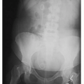
There is an accentuated forward upper thoracic curve, which may result in kyphosis (Fig. 8-1).
The senses of taste and smell decrease.
Assess the patient’s condition frequently; ask the patient whether he or she feels dizzy or as if they may fall. Ensure and protect the patient from injury during radiographic examinations. Assist and observe your patient as they are getting on and off the x-ray table at all times.
Implications for the Radiographer
Rapid changes in lighting, such as moving from a brightly lighted waiting room into a darkened examining room, may cause the elderly patient momentary blindness. Offer patients assistance so that they do not fall.
Loss of sense of smell and hearing loss must be considered. The radiographer must assess that the patient is able to hear directions. Speak loudly enough, and do not yell for the patient to understand what is being said. In addition, it is important to face the patient when speaking with him or her. Do not assume, however, that all older persons have a hearing deficit and need to be spoken to in an abnormally loud voice.
During fluoroscopic examinations, background noise from the equipment may prevent the patient from hearing the instructions. Be especially careful to clearly state instructions and check for understanding.
 FIGURE 8-1 Lateral chest radiographic image demonstrating kyphosis. |
Pulmonary System
Normal Changes of Aging
Pulmonary function changes with age; lung capacity diminishes owing to stiffening of the chest wall, among other changes.
The cough reflex becomes less effective.Related posts:

Stay updated, free articles. Join our Telegram channel

Full access? Get Clinical Tree