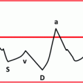
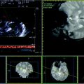
Fig. 8.1
Example of cerebral venous outflow measurement in supine position on the right side. a J3 IJV Doppler sampling and TAV measurement. b J3 IJV CSA measurement and automatic blood flow calculation in ml/min. c V1 VV Doppler sampling and TAV measurement. d V1 VV diameter measurement and automatic CSA and blood flow calculation
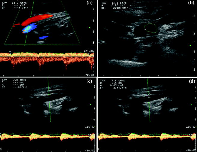
Fig. 8.2
Example of cerebral venous outflow measurement in supine position on the left side. a J3 IJV Doppler sampling and TAV measurement. b J3 IJV CSA measurement and automatic blood flow calculation in ml/min. c V1 VV Doppler sampling and TAV measurement. d V1 VV diameter measurement and automatic CSA and blood flow calculation
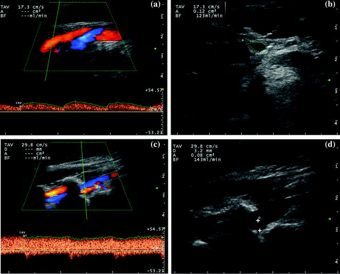
Fig. 8.3
Example of cerebral venous outflow measurement in upright position on the right side. a J3 IJV Doppler sampling and TAV measurement. b J3 IJV CSA measurement and automatic blood flow calculation in ml/min. c V1 VV Doppler sampling and TAV measurement. d V1 VV diameter measurement and automatic CSA and blood flow calculation
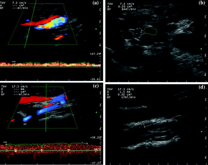
Fig. 8.4


Example of cerebral venous outflow measurement in upright position on the left side. a J3 IJV Doppler sampling and TAV measurement. b J3 IJV CSA measurement and automatic blood flow calculation in ml/min. c V1 VV Doppler sampling and TAV measurement. d V1 VV diameter measurement and automatic CSA and blood flow calculation
Related posts:
Stay updated, free articles. Join our Telegram channel
Full access? Get Clinical Tree
