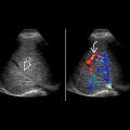
KEY FACTS
Terminology
- •
Gonadal stromal tumors arise from nongerm cell elements
Imaging
- •
Leydig cell tumors
- ○
Small, solid, hypoechoic intratesticular mass
- ○
May occasionally show cystic change
- ○
- •
Sertoli cell tumors
- ○
Small, hypoechoic mass with occasional hemorrhage, which may lead to heterogeneity and cystic components
- ○
± punctate calcification; large, calcified mass in large-cell calcifying Sertoli cell tumor
- ○
May produce estrogen/müllerian inhibiting factor
- ○
- •
Gonadoblastoma
- ○
Stromal tumor in conjunction with germ cell tumor, usually mixed sonographic features
- ○
Clinical Issues
- •
30% of patients with gonadal stromal tumors have endocrinopathy secondary to testosterone or estrogen production by tumor presenting with
- ○
Precocious virilization in children
- ○
Gynecomastia, impotence, ↓ libido in adults
- ○
- •
Majority of these tumors are benign
- •
Orchidectomy is preferred treatment
Diagnostic Checklist
- •
Consider stromal tumor in any patient with endocrinopathy and testicular mass
Scanning Tips
- •
May be indistinguishable from germ cell tumors on grayscale ultrasound but typically smaller in size
- •
High-frequency transducer (9-15 MHz) best imaging tool for detection of gonadal stromal neoplasms
 . The tumor is small and, like many sex cord stromal tumors, does not extensively involve the testis. Hemorrhage and necrosis are lacking.


