Chapter 2
Group 1: One or Two Clusters of Crushed Stone-like Calcifications on the Mammogram Produced by Malignant Processes
Group 1A. The mammographic finding represents the main morphological finding. In these cases there is concordance between mammography and the underlying histology. The temporal changes may be deceptively gradual. The outcome is excellent, even when invasion develops, provided the tumor is surgically removed when it is still in the 1-14 mm size range. The role of MRI in detecting these cases has yet to be defined.
Group 1B. The subtle mammographic finding may represent only the “tip of the iceberg.” In these cases the true extent of the disease is seriously underestimated by mammography, although it may be adequately outlined by functional imaging methods such as MRI, since the disease involves numerous TDLUs, ducts, and neoducts with little or no intervening normal tissue. The unexpectedly large tumor burden may result in a poor outcome, which can only become worse when underestimation of the disease leads to delayed or inadequate therapy.
Group 1A: The Mammographic Finding Represents the Main Histological Finding
Example 2.1
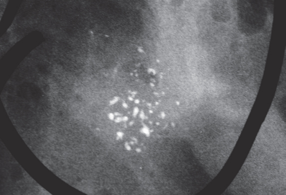
A 57-year-old asymptomatic woman, screening examination. Called back to the assessment center for further examination of the small cluster of calcifications in her left breast detected at screening.

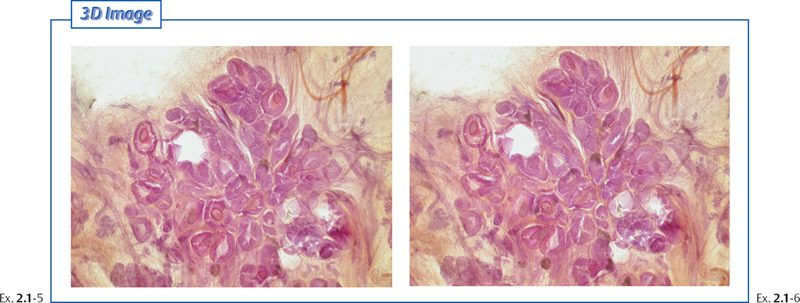
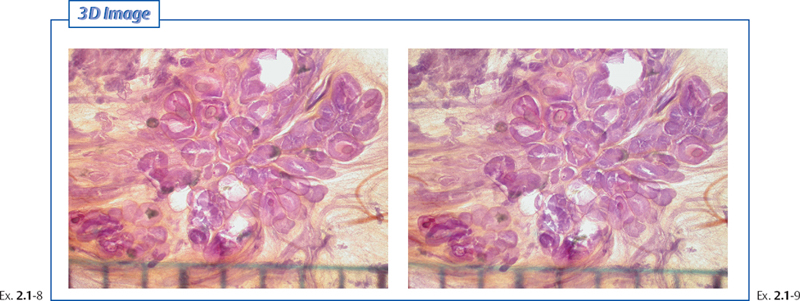
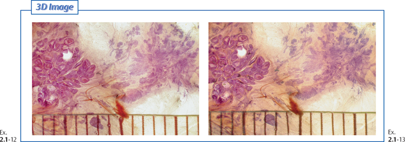
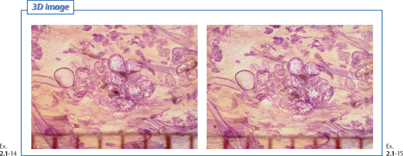
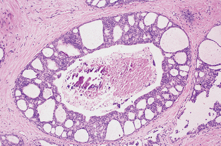
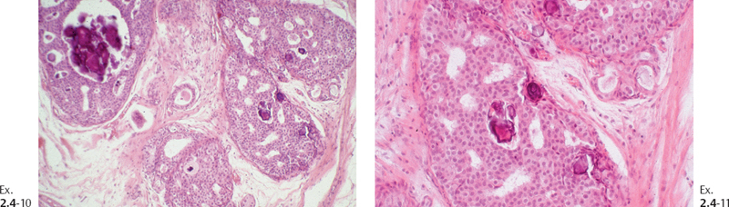
Ex. 2.1-12 & 13 Comparison of two adjacent TDLUs. The one on the left is altered by in situ carcinoma and the one on the right is deformed by sclerosing adenosis.
Treatment and follow-up: Wide surgical excision with no adjuvant therapy. The patient was free of disease at the most recent follow-up 13 years following surgery.
Example 2.2
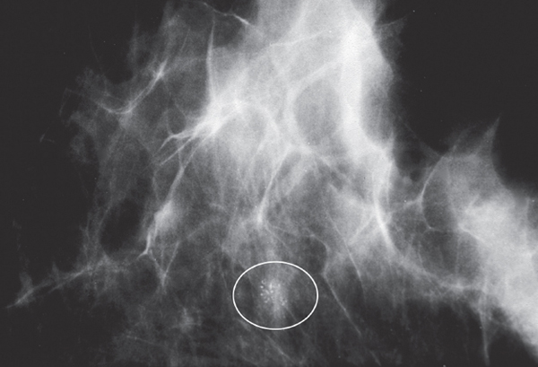
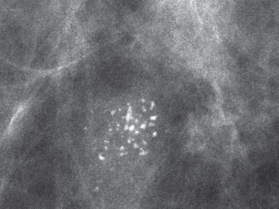
A 70-year-old asymptomatic woman, screening examination. The faint cluster of crushed stone-like calcifications was not perceived. At her next screening examination 18 months later she was still asymptomatic and was called back for further assessment of the cluster of calcifications.
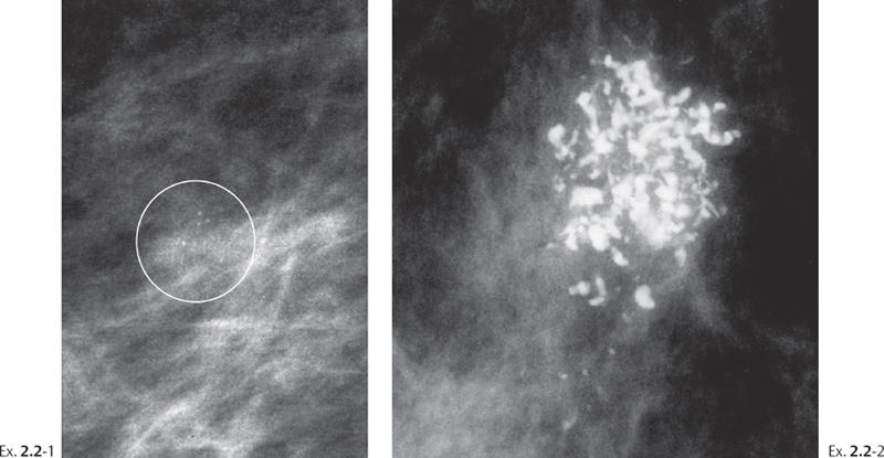
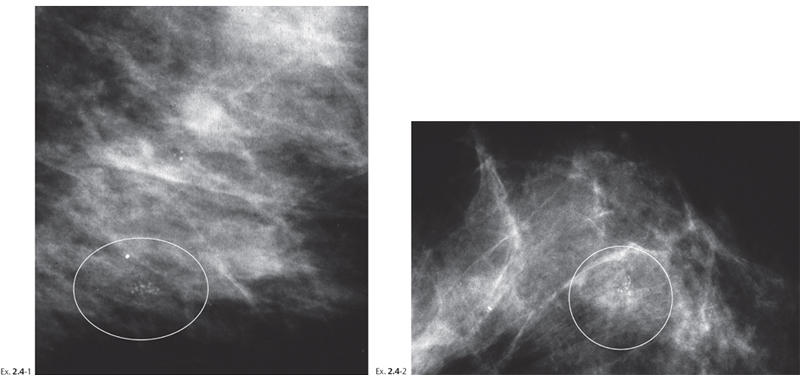
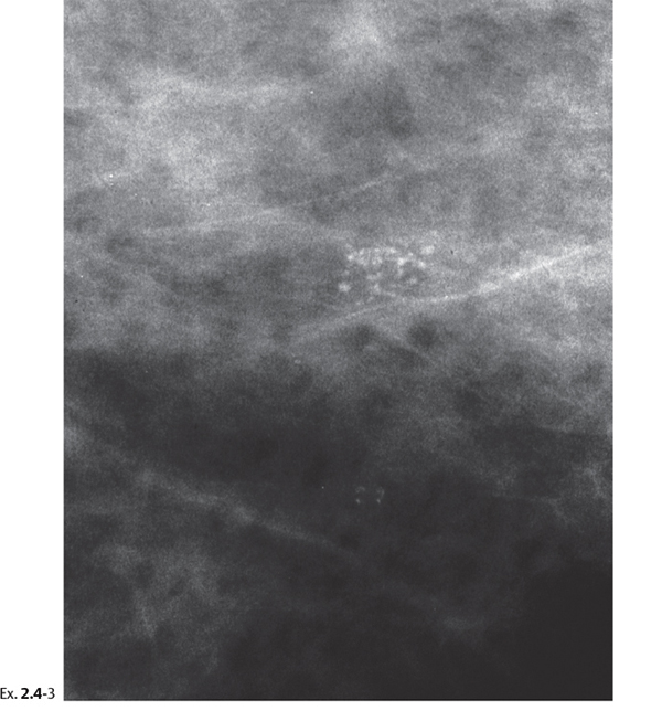
Ex. 2.2-1 & 2 Detail of the MLO projection in two consecutive screenings with an interval of 18 months. The barely perceptible cluster of discernible, crushed stone-like calcifications (within the circle) has evolved to a mixture of crushed stone-like and casting type calcifications, occupying a much larger volume.
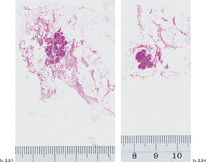
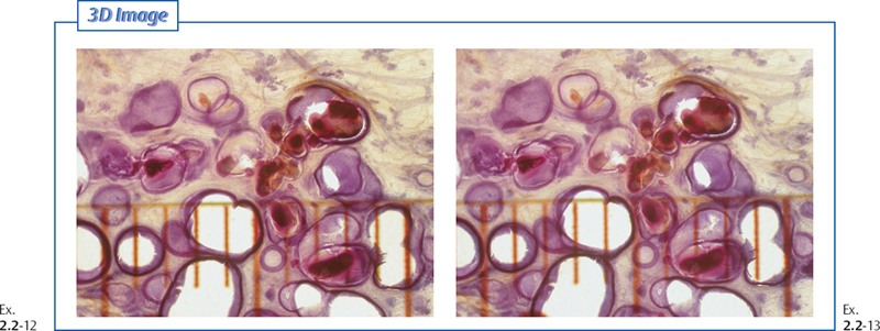
Histological diagnosis: 18 mm ⊠ 11 mm ⊠ 8 mm Grade 3 in-situ carcinoma with solid and cribriform cell architecture, containing multiple foci of microinvasion.
Treatment and follow-up: Sector resection was performed. No postoperative radiotherapy or other adjuvant treatment was given. The patient had no evidence of breast cancer at the most recent follow-up, 12 years after operation.
Comment
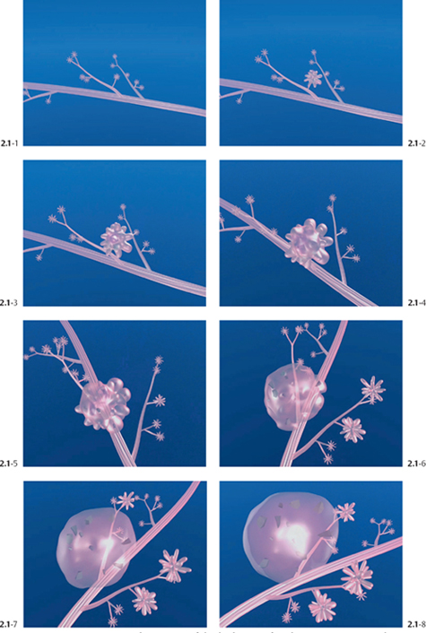
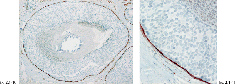
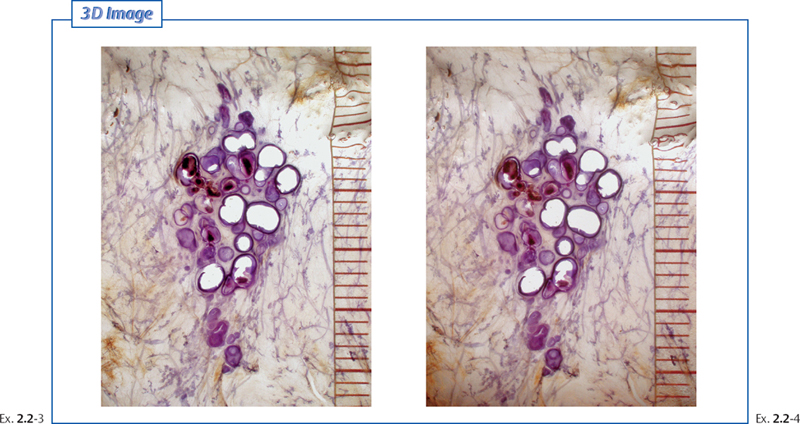
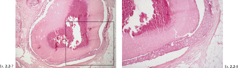
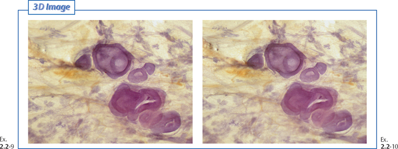
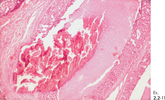
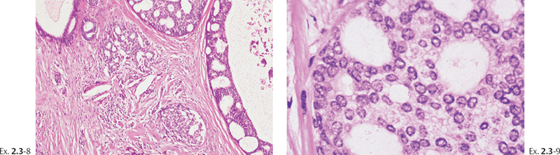
The large-section histological image and the subgross 3D images clearly show that the disease was limited to a single, extremely distended TDLU. The malignant cells were confined to I the acini of the lobule and no ductal involvement was found. These observations are at odds with the conventional term “ductal” carcinoma in situ. Sefton R. Wellings and his co-workers pointed out that pathologists, viewing traditional, small histological sections, might have mistaken the extremely distended acini for ducts.1 The fact that the disease is restricted to a single, isolated TDLU should be a justification for less radical treatment, such as surgical excision alone without adjunctive therapy.
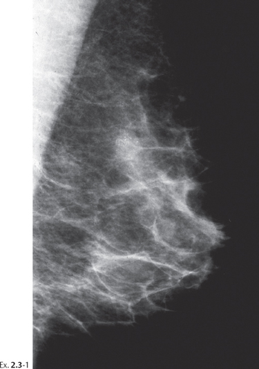
Example 2.3
A 64-year-old asymptomatic woman, screening examination.
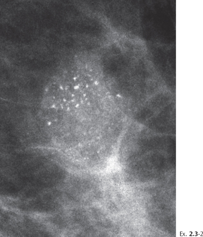
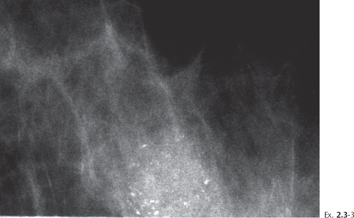
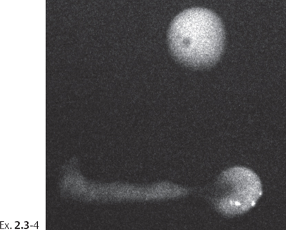
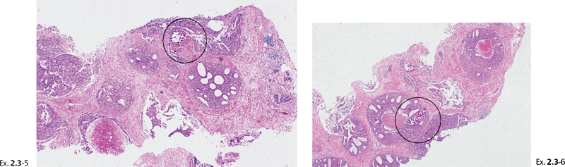
Treatment and outcome: Sector resection and postoperative irradiation. The patient had no evidence of recurrence at the most recent follow-up 9 years after treatment.
Example 2.4
A 58-year-old asymptomatic woman, screening examination.
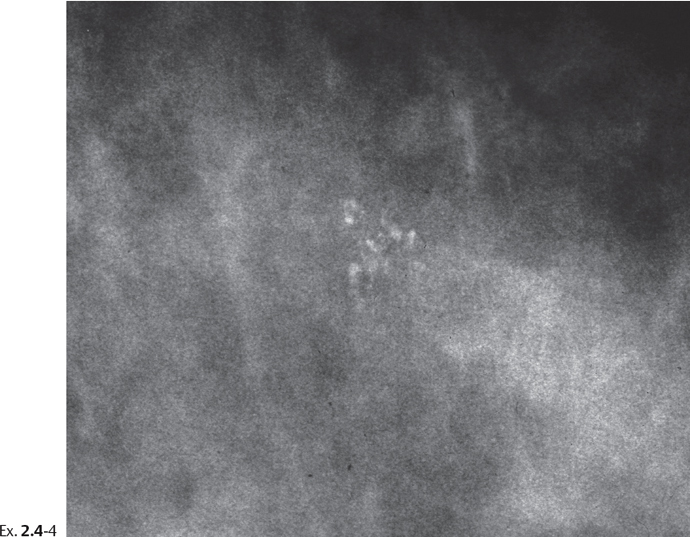
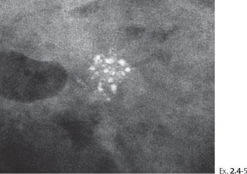
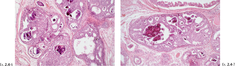
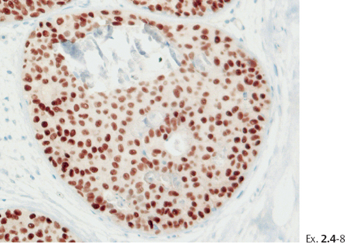
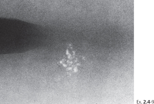
Treatment and outcome: Sector resection and postoperative irradiation. The patient was disease-free at the most recent follow-up examination, 14 years after treatment.
Example 2.5
A 52-year-old asymptomatic woman, first screening examination.
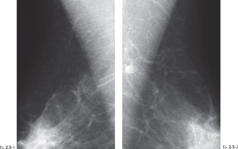
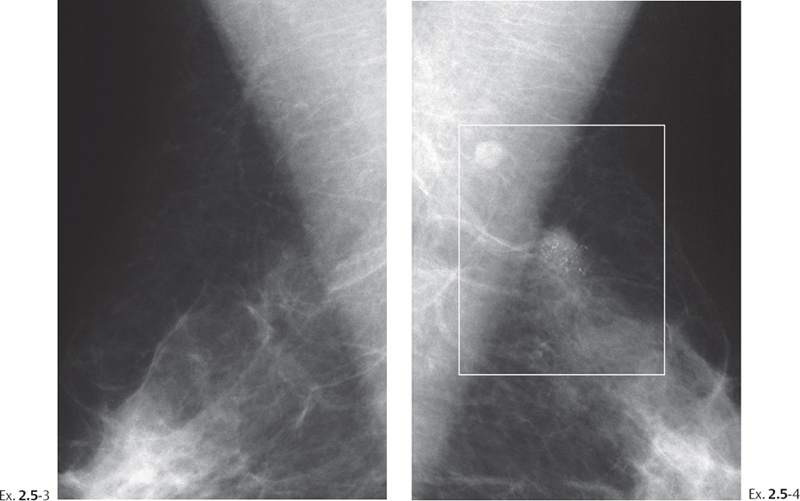
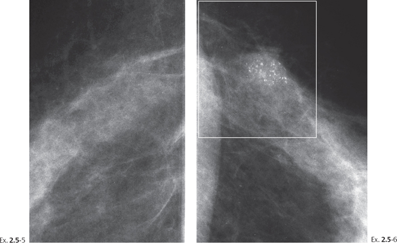
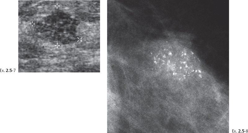
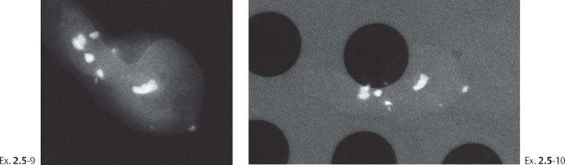
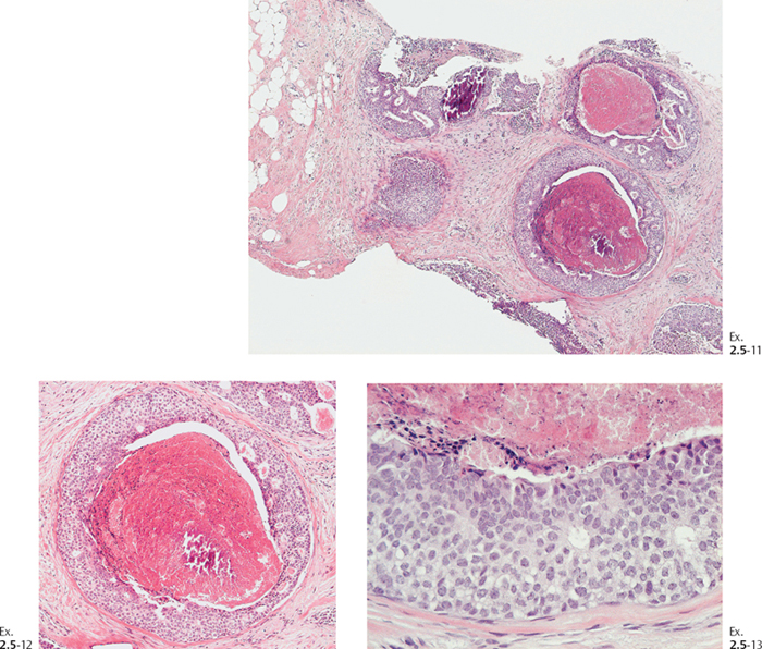
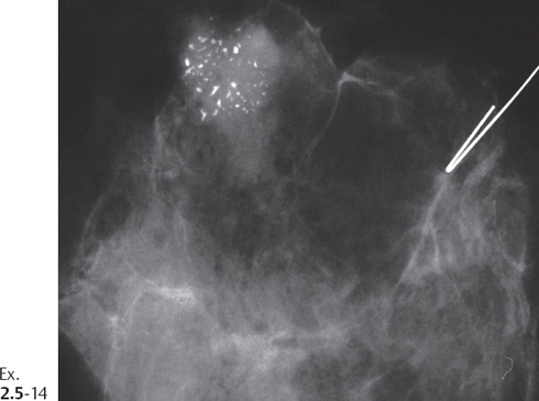
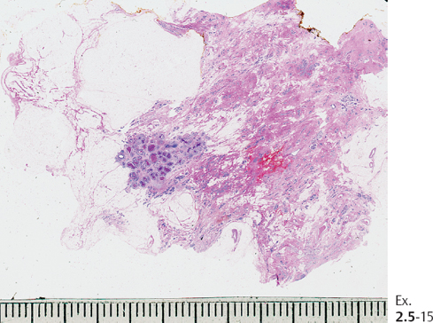
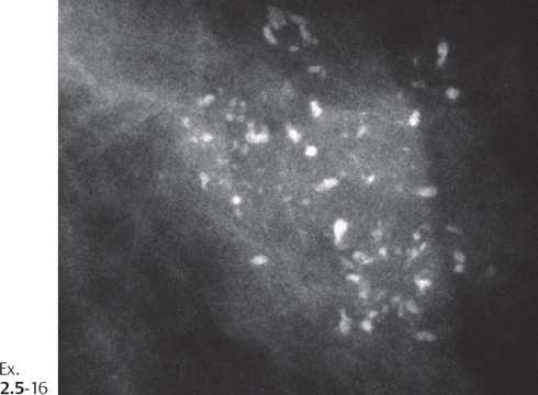
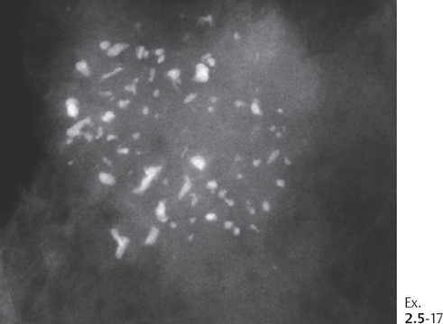
Treatment and outcome. Sector resection and postoperative irradiation. The patient had no sign of recurrence at the most recent follow-up examination 2 years after operation.
The Long-term Outcome of Cases in Group 1A
Regular mammographic screening offers us the opportunity of documenting the progression of the underlying disease in those occasional cases where the calcifications have been missed or misinterpreted as benign type and placed on follow-up, or where surgery has been delayed for other reasons. Consecutive mammographic examinations can document temporal changes in the appearance of the crushed stone-like calcifications (increasing or decreasing). These changes over time can help us learn more about the natural history of those breast cancer subtypes that develop within the TDLUs.
Two or more sequential mammographic examinations of Group 1A cases may reflect disease progression as follows:
- The mammographic image of crushed stone-like calcifications may show little apparent change at one or more follow-up examinations. In these cases short-term follow-up does not reliably exclude malignancy (see Example 1.1 on page 11).
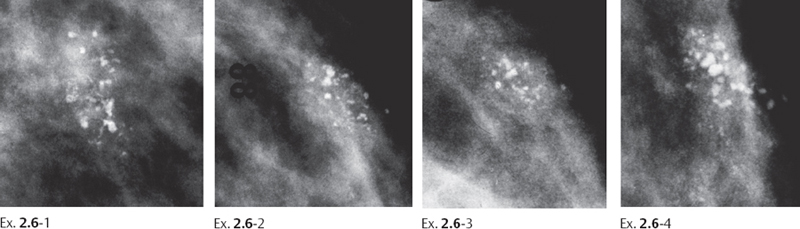
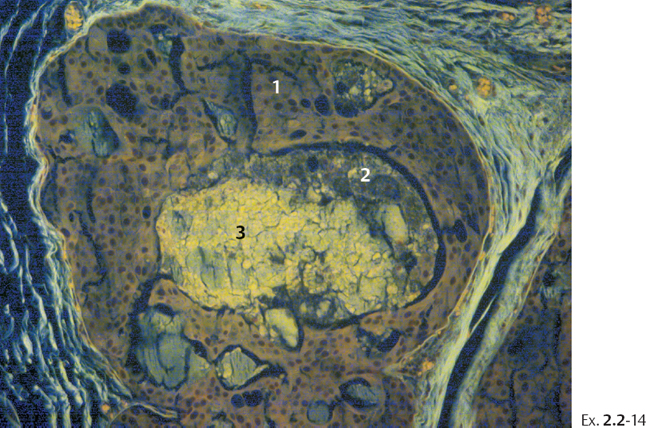
Ex. 2.6
Related posts:
 The Prognostic Importance of Mammographic Tumor Features
 The Diagnostic Approach to Malignant Type Calcifications on the Mammogram
 Differential Diagnosis of Breast Diseases Producing Clustered, Discernible Calcifications
 Multiple Clusters of Crushed Stone-like Calcifications on the Mammogram Produced by Malignant Processes
 Local Staging: Imaging Options and Core Biopsy Strategies
 Calcifications: Eggshell/Rim

Stay updated, free articles. Join our Telegram channel

Full access? Get Clinical Tree


