Worldwide, an estimated 10 million people are affected annually by traumatic brain injury (TBI). More than 5 million Americans currently live with long-term disability as a result of TBI and more than 1.5 million individuals sustain a new TBI each year. It has been predicted that TBI will become the third leading cause of death and disability in the world by the year 2020. This article outlines the classification of TBI, details the types of lesions encountered, and discusses the various imaging modalities available for the evaluation of TBI.
“No head injury is too trivial to ignore” —Hippocrates, 460–377 bc
The scope of the problem
Traumatic brain injury (TBI) is termed “the silent epidemic” for good reason. Worldwide, an estimated 10 million people are affected annually by TBI. In the United States alone, it is the leading cause of mortality and morbidity in individuals younger than 44 years, and its total direct and indirect cost to society has been estimated to exceed $60 billion annually. More than 5 million Americans currently live with long-term disability as a result of TBI and more than 1.5 million individuals sustain a new TBI each year. Furthermore, the number of victims continues to increase each year, and it has been predicted that TBI will become the third leading cause of death and disability in the world by the year 2020.
The primary etiology of head trauma varies with the age of the patient. Nonaccidental trauma (abuse) is most common in infants, whereas falls and sports-related injuries are seen in toddlers and school-aged children, respectively. Motor vehicle accidents are a frequent cause of head injury in young adults, but there is also increasing recognition of concussion in both amateur and professional athletes. The elderly population is particularly susceptible to accidental falls.
TBI classification
Clinically, TBI has been traditionally divided into minor, mild, moderate, and severe injury, as judged by the widely used Glasgow Coma Scale (minor: GCS = 15; mild: GCS ≥13; moderate: GCS 9–12; severe: GCS ≤8). Although the GCS has excellent interobserver reliability and correlation with outcome following severe brain injury, it fails to differentiate among different types of injury with different prognoses and fails to localize the injury. Of reported TBI cases, approximately 85% are classified as mild. It should be noted, however, that many cases of mild TBI go unreported. Unfortunately, mild TBI is the least well understood in terms of definition, imaging strategies, and correlation between imaging findings and long-term outcome. Routine structural imaging is often normal in these patients, yet studies have shown that even patients with normal imaging studies can have persistent cognitive deficits, and experience vocational and emotional distress that causes a significant socioeconomic burden to the individual and to society.
In addition to clinical severity, TBI can also be classified chronologically into primary and secondary injuries. Primary injuries are defined as those that occur at the moment of impact (eg, immediate tissue laceration by a gunshot injury). Secondary injuries are those that occur after the initial injury (eg, cerebral swelling and herniation, ischemia, venous thrombosis, posttraumatic infection, hydrocephalus, and cerebrospinal fluid leak) as a consequence of physiologic response to injury or complications of injury.
Whereas the primary injuries are considered irreversible, secondary injuries are potentially preventable with efficient triage and stabilization, management of parameters such as brain oxygenation, intracranial pressure, and cerebral perfusion pressure, and, in some cases, decompressive hemicraniectomy. This classification is somewhat arbitrary, however, because injury to the brain is actually more of a continuum of dynamically changing pathologies and not a single disease. For example, epidural hematoma (EDH) is classified as a primary injury, but the lesion does not necessarily present “at the moment” of injury, as it takes some time to expand. Brain injury triggers a cascade of pathophysiological events that can extend over a long period of time. Indeed, TBI should be thought of not as a static event, but rather a progressive injury with varying therapeutic windows.
TBI can also be classified according to the location of the injury (intra- and/or extra-axial) and the mechanism of the injury (penetrating/open, blunt/closed, or blast). The extra-axial lesions include epidural, subdural, subarachnoid, and intraventricular hemorrhages. The intra-axial lesions include the cortical contusion, intracerebral hematoma, traumatic axonal injury (TAI), and cerebrovascular injury. Each of these lesions is reviewed later in the section “Injuries”.
Imaging tools
The goals of neuroimaging are to identify treatable injuries, assist in the prevention of secondary damage, and provide useful prognostic information regarding the scope of a patient’s TBI.
Skull Radiography
Conventional plain film radiography of the skull in TBI has been supplanted by computed tomography (CT), but occasional indications include (1) to better define the position of penetrating objects and radiopaque foreign bodies, and (2) to better detect skull fractures in cases of suspected child abuse. In the vast majority of cases, however, the digital “scout” view of a CT scan, in combination with the CT data, is sufficient.
Computed Tomography
Noncontrast CT remains the study of choice for assessment of acute TBI because of its widespread availability, speed, safety, and compatibility with life-support and traction-stabilization devices, as well as its sensitivity to acute hemorrhage, hydrocephalus, herniation, fractures, and radiopaque foreign bodies. It is an excellent tool for deciding whether the patient should be surgically or medically triaged.
A relatively recent study showed that the cost of liberal CT screening for head trauma is justified because the consequences of undiagnosed brain injury can be devastating. Even with the tremendous advances in CT technology over the last 2 decades, however, most mild TBI cases show no visible abnormality on CT. Another downside of CT scanning is the radiation exposure, especially when multiple serial examinations in a young patient are necessary, as is frequently the case in the trauma setting.
Various clinical algorithms have been proposed to risk-stratify TBI patients for CT screening. In general, there is consensus that patients with moderate to severe intracranial injury (GCS ≤12) should undergo emergent noncontrast head CT. Several guidelines to determine whether patients with mild TBI (GCS >12) should undergo CT have been proposed, including the New Orleans Criteria and the Canadian CT Head Rule. The Canadian CT Head Rule proposed scanning patients with a GCS score of 13 to 15 based on these high-risk factors:
- •
Failure to reach a GCS of 15 within 2 hours
- •
Suspected open skull fracture
- •
Two or more vomiting episodes
- •
Signs of basal skull fracture
- •
Age older than 65.
CT angiography
CT angiography (CTA) uses intravenous iodinated contrast to delineate the vascular structures at sub-millimeter resolution. CTA is performed best with multidetector CT (MDCT) and rapid bolus contrast injection using vessel tracking. Typical imaging parameters include a slice thickness of 1.25 mm with a 0.625-mm overlap, and a bolus injection rate between 3 and 4 mL/s. CTA plays an important role in the diagnosis of suspected vascular injuries such as pseudoaneurysm, dissection, carotid cavernous fistula, or laceration-extravasation. CTA is usually the study of choice to screen for a possible cerebrovascular injury in the setting of penetrating injury or skull base fracture traversing the carotid canal or a venous sinus. If CTA is abnormal or suspicious, then conventional catheter angiography can be used to confirm and to potentially endovascularly treat the vascular injuries. Note that intravenous contrast should not be administered without a prior noncontrast examination, because intracranial contrast can both mask and mimic underlying hemorrhage.
CT perfusion
Energy consumption by the brain is high, accounting for roughly 20% of the oxygen and 25% of the glucose consumed by the body. In the uninjured brain, capillary blood flow to the brain is autonomously regulated (“autoregulation”). Insight into brain pathology can be gained through noninvasive imaging of cerebral perfusion and metabolism, and CT perfusion (CTP) is a technique that affords insight into the altered hemodynamics secondary to TBI. CTP can help clinicians distinguish between patients with preserved autoregulation and those with impaired autoregulation.
CTP is a bolus-tracking technique that involves continuous cine scanning through a limited volume of brain tissue with a scan interval of 1 second and a total scanning duration of 40 to 45 seconds. CTP measures the transient attenuation changes in the blood vessels during the first pass of an intravenously injected contrast bolus. Color maps of cerebral blood volume (CBV), mean transit time (MTT), and cerebral blood flow (CBF) are then generated from these time-attenuation curves and can be used to assess cerebral perfusion.
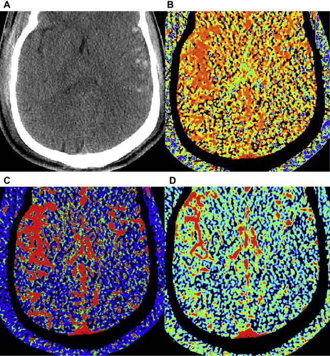
Although CTP is fairly routine in stroke and brain tumor imaging, it is rarely performed in TBI. Nevertheless, research studies have shown that CTP can be used to demarcate acute focal brain injury, document diffuse injury, identify areas at risk of secondary injury, and predict enlarging contusions. Visible changes in CBV on either CTP or perfusion magnetic resonance (MR) imaging have been shown to occur in areas of contusion visible on conventional CT or MR imaging ( Fig. 1 ). CTP can provide prognostic information regarding outcome, with normal brain perfusion or hyperemia in patients with favorable outcome and oligemia in patients with unfavorable outcome. Controversy exists, however, regarding correct selection of an arterial input vessel, the accuracy of quantitative results, and the reproducibility of results. Other limitations include CTP’s limited anatomic coverage, and the additional radiation and contrast exposure that accompany this method.
Dual-energy CT
Newer CT technologies that allow for more rapid data acquisition have sparked renewed interest in dual-energy application. Knowing how a substance (eg, blood) behaves at 2 different energies can provide information about tissue composition beyond that obtainable with single-energy techniques. The current applications of dual-energy CT in head injury are unknown, but in the future are likely to include providing information about tissue structure, predicting the evolution of soft tissue injuries, the ability to generate “virtual” unenhanced images, and the improved detection of iodine-containing substances on low-energy images, thereby improving CTA and CTP in head injury at a reduced radiation dose.
Magnetic Resonance Imaging
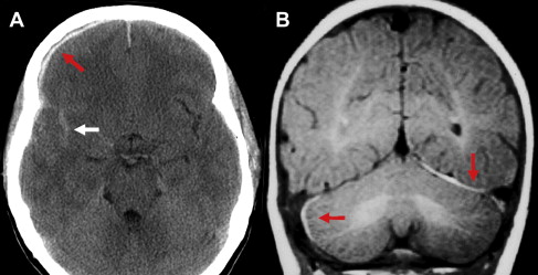
On occasion, MR imaging may be indicated in patients with acute TBI if the patient’s neurologic findings are unexplained by CT findings. MR imaging is also helpful in cases of suspected nonaccidental trauma within the first few days of injury, as it is far superior to CT in the detection of TAI and small subdural “smear” collections that might be the only clue to child abuse ( Fig. 2 ). In general, however, MR imaging is limited in the emergency setting due to safety considerations and its relatively long imaging times; imaging time can be a significant limitation in some patients, especially if they are uncooperative, claustrophobic, or medically unstable. Furthermore, MR’s incompatibility with many trauma-related or other medical devices, its relative insensitivity to acute subarachnoid hemorrhage, and its sensitivity to patient motion make it more easily and appropriately performed for assessment of subacute and chronic TBI. MR imaging can also be used as a problem-solving tool in the setting of neurologic deficits unexplained by CT; MR may identify brainstem injury, ischemia/infarction, small cortical contusions, and white matter injury, all of which are relative “blind spots” on CT.
The key MR imaging sequences to perform include: fluid-attenuated inversion recovery (FLAIR) images for nonhemorrhagic TAI and subarachnoid hemorrhage (SAH); gradient-recalled echo (GRE) or susceptibility-weighted imaging (SWI) for hemorrhagic TAI and blood products in general; and diffusion-weighted imaging (DWI) for ischemia/infarction and TAI. These sequences are now discussed, followed by a brief discussion of more advanced MR imaging techniques.
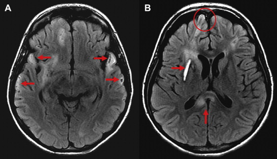
FLAIR is a T2-weighted inversion recovery sequence that suppresses the T2 bright signal from cerebrospinal fluid (CSF), thus improving lesion conspicuity in the periventricular regions and cerebral cortex. FLAIR is far more sensitive than conventional T2-weighted images to cortical contusions, TAI, and SAH ( Figs. 3–6 ).
GRE T2*-weighted imaging is sensitive to the presence of blood breakdown products: deoxyhemoglobin, intracellular (not extracellular) methemoglobin, ferritin, and hemosiderin. These blood products results in areas of signal loss due to alteration of the local magnetic susceptibility in the tissue ( Fig. 7 ). Because hemosiderin can persist indefinitely, GRE T2*-weighted imaging is recommended for the evaluation of remote TBI as well as for acute and subacute TBI. Small foci of hemosiderin can sometimes be resorbed over time, however, so the lack of evidence of blood products on GRE does not exclude prior TBI.
Limitations of GRE images include geometric distortion, chemical-shift artifacts, and susceptibility artifacts at bone-air-brain interfaces (especially over the frontal and ethmoidal sinuses and mastoid air cells) that obscure the anatomy of the orbitofrontal and inferior temporal lobes, both areas that are typically injured in TBI ( Fig. 8 ). The choice of echo time (TE) affects GRE artifacts and performance: whereas a short TE reduces susceptibility effects at the tissue-air boundaries, a longer TE sequence (which maximizes lesion-to-tissue contrast) more sensitively detects TAI lesions. Fortunately, these geometric distortions and susceptibility effects can be mitigated so some extent by parallel imaging and fast image acquisition methods.
SWI can be likened to a supercharged GRE sequence and is the most sensitive sequence for identifying hemorrhage, especially petechial hemorrhage. SWI amplifies susceptibility effects by combining both magnitude and phase information from a high-resolution, fully velocity compensated 3-dimensional (3D) T2*-weighted gradient-echo sequence. Conventional GRE T2*-weighted MR imaging relies only on the magnitude images and ignores the phase images, the latter of which contain valuable information regarding tissue susceptibility differences. SWI is 3 to 6 times more sensitive than GRE T2*-weighted imaging in detecting hemorrhagic TAI. In addition, a recent study by Wu and colleagues showed that it is also fairly sensitive to acute SAH as well as subacute and chronic SAH.
DWI is one of the imaging sequences of choice for acute TAI and ischemia/infarction, but it is also helpful in identifying cerebral contusions. In the central nervous system, the diffusion of water is impeded by tissue structures such as cell membranes, myelin sheaths, intracellular microtubules, and associated proteins. Because living cells and tissues are composed of multiple subcompartments where the magnitude and direction of diffusion vary, diffusion is faster in the extracellular space than in the intracellular space. As in cerebral infarction, acute TAI lesions also show increased signal (reduced diffusion) on DWI with a corresponding reduction in apparent diffusion coefficient (ADC). DWI generally reveals more lesions than fast spin-echo T2-weighted and GRE T2*-weighted images in patients imaged within 48 hours of injury ( Fig. 9 ). Chronic TAI lesions are either invisible or show decreased signal (facilitated diffusion) on DWI.
Diffusion tensor imaging (DTI) is sensitive to the spatial orientation of water diffusion, and allows for the virtual reconstruction of axonal networks within the brain. DTI has provided important insights into the neurobiological basis for normal development and aging, as well as various disease processes in the central nervous system such as demyelinating disorders, brain tumors, dysmyelination, and TBI. DTI is the study of choice when evaluating the integrity and directionality of white matter fiber tracts, and thus it has been extensively applied as a research tool in the study of TAI.
There are 2 primary indices of DTI: fractional anisotropy (FA) and ADC. FA is an index of water diffusivity in the voxel. FA is low when fibers are not collinear (eg, crossing fibers) or if fibers have been damaged. ADC is an estimate of the average magnitude of water movement in a voxel. Therefore, ADC will increase in the setting of vasogenic edema and when water flows out of capillaries into the interstitial space. ADC decreases with cytotoxic edema and when diffusion is restricted by injured swollen cells. When axons are injured, normal anisotropy decreases because of restricted axoplasmic flow. Several studies have revealed an abnormal decrease in white matter FA when conventional MR imaging was normal. A follow-up study in 2 patients with TBI revealed that in some regions with initially reduced FA, the changes were partially or completely corrected 30 days after injury. These results should not necessarily be interpreted as evidence for regeneration of neurons. Instead, a cellular repair mechanism may have corrected the cytoskeletal misalignment before disconnection occurred.
DTI is providing some remarkable insights into TBI, but there appears to be an “irrational exuberance” in the literature, especially for 3D-color tractography ( Fig. 10 ). The sensitivity of fiber-tracking algorithms to many physical and computational variables is still poorly understood, and their behavior in the face of injured tissue even less so. Numerous issues must still be accounted for to reduce false positives and false negatives when interpreting DTI studies. For example, FA and ADC are affected by imaging variables such as field strength and resolution, as well as patient variables including age and preexisting disease. Although single-shot echo-planar imaging (EPI) is fairly immune to extreme variability in phase between applications of diffusion-encoding gradients, it suffers from severe artifacts in the presence of magnetic field inhomogeneities. The significant T2* decay that can occur during long echo trains makes high-resolution acquisitions challenging.
With the recent advance of parallel imaging technology, less spatial distortion and higher signal-to-noise ratio can be achieved in a time-efficient manner with single-shot EPI. More case-control studies are necessary, however, with age-matched comparisons of DTI results between patients and corresponding control subgroups. Finally, the difficulties of post-processing and the need for statistical analyses to evaluate the effects on FA maps of spatial distortion correction induced by gradient nonlinearity, misregistration, image processing, and variable protocol parameters (b value, number of diffusion-encoding gradient directions, number of excitations, software data analysis package, and so forth) are currently keeping DTI primarily in the research arena.
Magnetization transfer imaging (MTI) takes advantage of the molecular disarrangement of white matter tracts in TAI. MTI exploits the longitudinal (T1) relaxation coupling between bound (hydrated) water protons and free (bulk) water protons. Although classically used to assess demyelination in the setting of multiple sclerosis, MTI may provide a quantitative index of the structural integrity of tissue, and therefore is a potential marker of TAI.
Magnetic resonance spectroscopy (MRS) can reveal posttraumatic neurometabolite abnormalities in the injured brain when conventional neuroimaging is normal. The most common brain metabolites that are measured with proton [ 1 H]MRS include N -acetylaspartate (NAA), creatinine (Cr), choline (Cho), glutamate (Glu), lactate, and myoinositol. In brief, a reduction of NAA, a marker of axonal and neuronal functional status, has been shown to be a dynamic process after TBI: it remains low in patients with poor recovery and returns to normal in patients with good outcomes. Decreased NAA values represent either neuronal loss or neuronal/mitochondrial dysfunction, because they can recover after the resolution of energy failure. Total Cho is considered an indicator of membrane structural integrity, and is typically increased in TBI. Ross and colleagues suggested that elevated Cho levels in white matter may be caused by breakdown products appearing after the shearing of myelin and cellular membranes, and that reduced NAA values result from axonal injury. Changes in Cr, a marker of cell energy metabolism and mitochondrial function, may also be seen. An increased Cr level may be part of a repair mechanism associated with increased mitochondrial function in areas of injury. Perturbations in Glu, the brain’s major neurotransmitter, as well as an increase in lactate are also seen following TBI. MRS is not routinely performed in TBI and is usually reserved for experimental studies; this is partially because the data interpretation is complicated by the variability in results depending on the severity of the initial injury, the time after injury that the scan is performed, the types and location of spectral acquisitions, and the outcome measures used to monitor recovery. In addition, results may have been influenced by technical factors involved in the acquisition and processing of MRS data. One cannot account for voxels with spectra that are too distorted or that contain no measurable metabolites because of large amounts of blood products. This latter limitation may cause an underestimation of the overall effect of hemorrhagic damage. Finally, because ratios are often used instead of quantitative metabolite levels, changes in Cr may affect the ratios and may explain why the Cho/Cr ratio is slightly lower in patients with poor outcomes in some regions.
Functional MR imaging ( fMRI ) indirectly observes the activity of the brain via detection of an alteration in the ratio of cerebral blood deoxyhemoglobin to oxyhemoglobin in response to particular tasks. fMRI relies on the principle that changes in the blood oxygen level dependent (BOLD) signal are caused by changes in CBF, which in turn is thought to be caused by changes in neuronal activity. As neuronal activity increases, blood flow overcompensates such that the local blood oxygenation actually increases. Because deoxyhemoglobin is an endogenous paramagnetic contrast agent, a decrease in its concentration is reflected as an increase in signal intensity on GRE images. This principle has been validated in animal studies, but the fundamental mechanisms underlying the coupling between neuronal activity and blood flow changes have not been elucidated. This aspect is relevant to the topic of TBI because it is possible that abnormalities in fMRI signals could be caused by abnormalities in neuronal activity (as is usually assumed), or by abnormalities in the coupling of neuronal activity to the regulation of CBF (which is rarely assessed).
fMRI can be performed at either 1.5 T or 3 T, but higher field strength is generally preferred. A high-resolution 3D-SPGR (spoiled gradient-recalled acquisition in the steady state), T1-weighted whole brain study is initially obtained. Then, for fMRI data collection, GRE EPI is performed, preferably with parallel imaging. Although “resting state” fMRI can be performed, most fMRI examinations measure the magnetic evoked response to a stimulated task that is subsequently coregistered with the high-resolution MR images.
fMRI research in TBI has centered primarily on the assessment of deficits after mild TBI ( Fig. 11 ), but also offers promise in the understanding of the brain’s ability to reorganize after injury. As with the aforementioned advanced MR imaging techniques, however, fMRI has been used in number of research studies of TBI patients but is not yet part of routine clinical care. There is a need for improved standardization of fMRI acquisition and analysis protocols for TBI. A major challenge facing task-related fMR imaging studies is that the changes in BOLD signal are strongly dependent on how well the task is performed, so interpretation of whether fMRI changes are caused by TBI or simply by worse performance is a major issue. Another criticism of fMRI in TBI is the lack of a baseline (pre-trauma) study.
Voxel-based morphometry (VBM) measures the change in volume of the brain and has traditionally been used to assess brain atrophy in Alzheimer disease. Only preliminary research in TBI patients has been performed, but gray matter atrophy has been identified, suggesting that the eventual pathologic end point of TBI is loss of cortical neuronal cell bodies. Two possible mechanisms are thought to be responsible. First, the injury could affect cortical structures as the brain impacts the cranial vault in a coup-contrecoup manner. Second, TAI could cause axonal damage, leading to retrograde degeneration and neuronal somatic loss. Postsynaptic cortical atrophy may also result from diminished anterograde transmission.
Single-photon emission tomography (SPECT) uses the gamma-emitting isotope technetium-99 to image cerebral perfusion. The discovery of significant changes in CBF in patients with TBI makes SPECT a promising tool in evaluating patients with mild TBI. Frontal and temporal lobe hypoperfusion is commonly seen in head injury, presumably due to the gliding effect of the brain over the underlying skull. Although this hypoperfusion is generally attributed to the cerebral edema that surrounds the damaged brain and limits CBF, it may also result from vasospasm, direct vascular injury, and/or perfusion changes due to alterations in remote neuronal activity (diaschisis). SPECT can show areas of perfusion abnormality following head trauma that are normal on conventional CT and MR imaging. Due to its low spatial resolution, however, SPECT is less sensitive in detecting many smaller lesions that are visible on MR imaging.
Positron emission tomography (PET) uses [ 18 F]2-fluoro-2-deoxy- d -glucose ( 18 F-FDG) to evaluate cerebral glucose metabolism in vivo. Acutely injured brain cells show increased glucose metabolism following severe TBI because of intracellular ionic perturbation. Following the initial hyperglycolysis state, injured brain cells then show a prolonged period of regional hypometabolism. Based on the principle that regional glucose metabolism reflects the neuronal activity of the region, focal hypometabolism indicates an area of neuronal dysfunction. To account for the metabolic reduction, 2 main mechanisms have been proposed: (1) local neuronal loss and (2) decreased neuronal activity as a result of deafferentation. FDG-PET also has an advantage for elucidating focal brain dysfunction compared with the cerebral perfusion information that is obtained from SPECT studies. Although a significant number of functional imaging studies in TBI have been performed using perfusion SPECT analysis, metabolic PET technology demonstrates similar overall findings but with increased resolution. Unfortunately, although PET provides information on cerebral metabolism and has shown promise in TBI, it is not widely available, is expensive, and is not regularly reimbursed for the evaluation of TBI.
Magnetoencephalography
Magnetoencephalography (MEG) is a noninvasive functional imaging technique with high temporal resolution (<1 millisecond) and spatial localization accuracy at the cortical level (2–3 mm). Unlike normal spontaneous MEG data, which are dominated by neuronal activity with frequencies above 8 Hz, brain-injured tissue generates abnormal low-frequency delta-wave (1–4 Hz) magnetic signals that can be directly measured and localized using MEG. Abnormal focal slowing can be detected by MEG when other imaging modalities are normal, and MEG appears to be more sensitive than MR imaging and SPECT to TBI. Abnormal MEG delta waves have been observed in subjects without obvious DTI abnormality, indicating that MEG may also be more sensitive than DTI in diagnosing mild TBI. MEG’s availability is unfortunately, very limited, with only about 20 whole-head MEG systems in the United States and fewer than 150 systems installed worldwide. This lack of availability will likely relegate MEG to the research arena for the foreseeable future.
The Injuries
Extra-Axial Injury
Scalp and skull injury
The scalp is composed of 5 layers: skin; subcutaneous fibro-fatty tissue; galea aponeurotica; loose areolar connective tissue; periosteum.
The subgaleal hematoma is a frequent sequela of head injury resulting from direct impact or shearing of traversing veins. Close examination of the scalp soft tissues should be one of the first steps in interpreting a trauma head CT. Identification of soft tissue injury allows the radiologist to pinpoint the site of impact, or “coup” site, that may be invisible to the clinician because of its small size or obscuration by the patient’s hair. Indeed, the presence of a scalp injury may be the only clue to the presence of TBI in a patient presenting with “altered mental status.”
The coup site in particular should be carefully inspected for the presence of an underlying skull fracture, as well as for evidence of soft tissue laceration and radiopaque foreign bodies. The 3 main types of skull fractures include linear (most common), depressed, and basilar. Fractures can also be described as comminuted (multiple fragments) or compound (open).
Linear fractures in the axial plane can easily be missed on axial CT slices, but close examination of the “scout” image will often reveal the fracture line ( Fig. 12 ).


