Hepatic TB and Fungal Infections
Michael P. Federle, MD, FACR
Brooke R. Jeffrey, MD
Key Facts
Terminology
Opportunistic infection of the liver (± other viscera), usually by fungal or mycobacterial organisms
Imaging
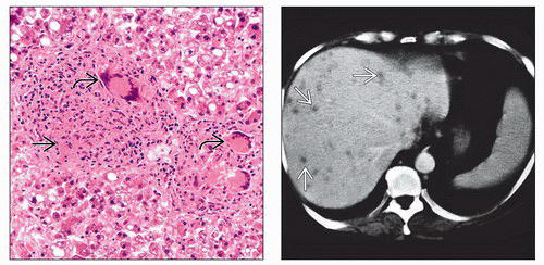
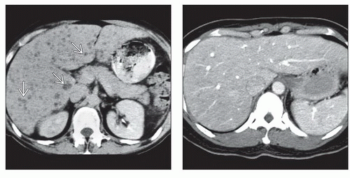
Best diagnostic clue: Multiple well-defined, rounded microabscesses in liver
Best imaging tool: CECT, US
Protocol advice
MDCT: IV contrast at 2.5 mL/sec with < 2 mm collimation and 2.5 mm reconstruction interval
MR: FLASH sequences
Gadolinium enhancement required to detect very small lesions
Top Differential Diagnoses
Metastases
Lymphomatous/leukemic foci in liver
Biliary hamartomas
Caroli disease
Pathology
Originates from intestinal seeding of portal and venous circulation
Clinical Issues
Most common signs/symptoms
Asymptomatic or abdominal pain and fever
Erythematous papules on skin
Clinical profile: Immunocompromised patient recovering from neutropenia
Diagnostic Checklist
Rule out other “innumerable hypodense liver lesions”
Biopsy specimen for histology/microbiology
TERMINOLOGY
Definitions
Opportunistic infection of liver (± other viscera), usually by fungal or mycobacterial organisms
IMAGING
General Features
Best diagnostic clue
Multiple well-defined, rounded microabscesses in liver
CT Findings
NECT
Multiple small, hypodense lesions
± scattered areas of calcific density (healing phase)
CECT
Biphasic CT may be more accurate than venous phase only
Nonenhancing hypodense centers
± peripheral rim enhancement
Central or eccentric “dot” felt to represent hyphae
Ultrasonographic Findings
Grayscale ultrasound
4 major patterns of hepatic candidiasis
Uniformly hypoechoic
Most common appearance (fibrosis and debris)
Echogenic
Scar formation
“Wheel within wheel”
Peripheral zone surrounds inner echogenic “wheel,” which surrounds central hypoechoic nidus (early stage)
“Bull’s eye”
1-4 mm lesion with hyperechoic center surrounded by hypoechoic rim (neutrophil count returns to normal)
After antifungal therapy: Lesions increase in echogenicity, decrease in size, often disappear altogether
Imaging Recommendations


