Hepatomegaly
Michael P. Federle, MD, FACR
Brooke R. Jeffrey, MD
Key Facts
Terminology
Enlargement of liver due to underlying pathologic process
Imaging
Best diagnostic clue: Hepatic length > 15 cm
Normal liver length 8-12 cm for men, 6-10 cm for women in midclavicular line
Liver protrudes beyond costal margin
Best imaging tool: Longitudinal US or coronal triphasic CT
Top Differential Diagnoses
Riedel lobe
Caudate hypertrophy in Budd-Chiari or PSC
Liver displacement by COPD
Lobar regeneration with major liver resection
Clinical Issues
Most common signs/symptoms
Palpable liver edge below costal margin
Other signs/symptoms
Jaundice, RUQ pain, fever, abnormal liver function tests
Benign outcome to fulminant liver failure
Often biopsy required for definitive diagnosis
Diagnostic Checklist
Hepatitis, steatosis, passive congestion, and diffuse tumor are most common causes
Image interpretation pearls
Consider triphasic CT to detect and characterize mass lesions
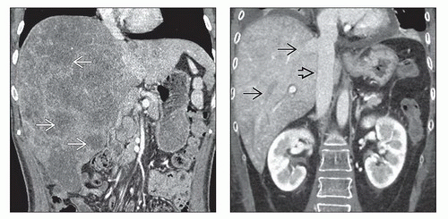 (Left) Coronal CECT reformation of a 52-year-old man with metastatic colon cancer shows marked enlargement of the liver with diffuse metastases throughout the right lobe  . (Right) Venous phase coronal CECT of a 27-year-old woman with leukemia and new onset of RUQ pain shows an enlarged and heterogeneous liver with distended hepatic veins . (Right) Venous phase coronal CECT of a 27-year-old woman with leukemia and new onset of RUQ pain shows an enlarged and heterogeneous liver with distended hepatic veins  and IVC and IVC  due to passive congestion of the liver. Drug toxicity accounted for the cardiac dysfunction. due to passive congestion of the liver. Drug toxicity accounted for the cardiac dysfunction. |
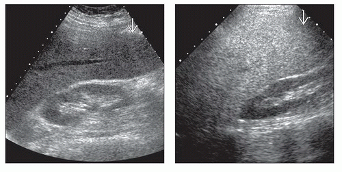 (Left) Longitudinal ultrasound in a 44-year-old man with a history of hepatitis B infection shows marked enlargement of the right lobe of the liver, extending caudally below the level of the kidney
 . (Right) Longitudinal ultrasound in a 38-year-old woman with nonalcoholic steatohepatitis reveals marked hepatic enlargement. Note the right lobe extending below the level of the right kidney . (Right) Longitudinal ultrasound in a 38-year-old woman with nonalcoholic steatohepatitis reveals marked hepatic enlargement. Note the right lobe extending below the level of the right kidney  as well as echogenicity greater than that of the kidney due to diffuse steatosis. as well as echogenicity greater than that of the kidney due to diffuse steatosis.Related posts:Stay updated, free articles. Join our Telegram channel
Full access? Get Clinical Tree
 Get Clinical Tree app for offline access

|