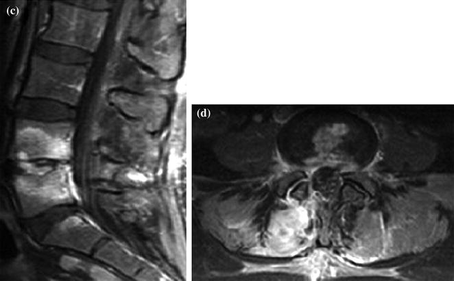
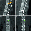
Fig. 1
a–d. SE T1 (a), FSE T2 fat sat sagittal (b); CE fat sat SE T1 sagittal (c) and axial (d). L4–L5 infective spondylodiscitis with involvement of intervertebral disk and bodies which are characterized by typical signal alteration (hyper intensity in T2–b) and CE (c–d)
< div class='tao-gold-member'>
Only gold members can continue reading. Log In or Register to continue
Related posts:
 Herniated Lumbar Disk Diskectomy
 Dorsal Herniated Disk Diskectomy and Stabilization
 Lumbar Stenosis and Degenerative Instability Posterior Rigid Stabilization
 Traumatic Lumbar Collapse Rigid Stabilization
 Lumbar Collapse in Chordoma Vertebral Drawing
 Dorsal Collapse in Multiple Myeloma Vertebroplasty
Stay updated, free articles. Join our Telegram channel

Full access? Get Clinical Tree


