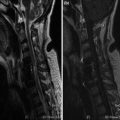
Fig. 1
a–g. FSE T2 sagittal (a–c) and axial (d–g) sections. Small right intraforaminal hernia, (arrow) in L3–L4 compressing the adjacent nerve root
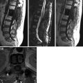
Postoperative Follow-Up After 1 Month
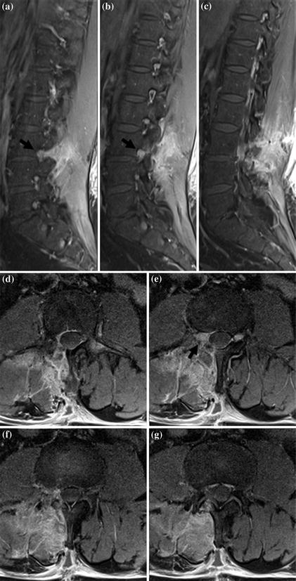


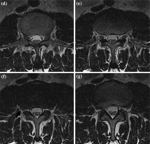
Fig. 2
a–g. CE fat sat SE T1 sagittal and axial sections. Exuberant granulation tissue characterized by intense CE, in the right retrovertebral space at L3–L4. a–g. This tissue extends over lamino-flavectomy in the right foramen, close to L3 nerve root, thickened by flogosis (arrow)
< div class='tao-gold-member'>
Only gold members can continue reading. Log In or Register to continue
Related posts:
Stay updated, free articles. Join our Telegram channel
Full access? Get Clinical Tree
