Fig. 1
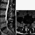
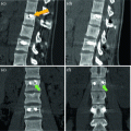
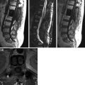
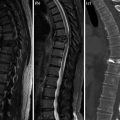
a–c. Sagittal CE SE T1 (a), sagittal CE fat sat SE T1 (b–c). Slight physiological CE (non-infectious) of subchondral spongiosa at L3–L4 for the presence of reactive granulation tissue, rear disk profile close to the annulus is also involved (a). These findings are emphasized in fat sat imaging (b–c). Regular CE of para-spinal soft tissue at the surgical breach (laminectomy). Regular CE is also appreciable at the disk L1–L2 where coexists ernia intraspongiosa with the same CE (aseptic discitis)
< div class='tao-gold-member'>
Only gold members can continue reading. Log In or Register to continue
Stay updated, free articles. Join our Telegram channel

Full access? Get Clinical Tree


