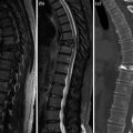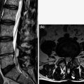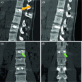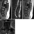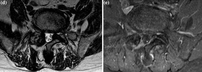
Fig. 1 a–e. FSE T1 sagittal (a), FSE T2 sagittal (b) and axial (d), CE fat sat T1 sagital (c) and axial (e)< div class='tao-gold-member'> Only gold members can continue reading. Log In or Register a > to continue Share this:Click to share on Twitter (Opens in new window)Click to share on Facebook (Opens in new window) Related posts: Herniated Lumbar Disk Micro-Diskectomy Recurrent Herniated Lumbar Disk Patient Reoperated Traumatic Lumbar Dislocation Percutaneous Stabilization Traumatic Lumbar Collapse Rigid Stabilization Lumbar Collapse in Chordoma Vertebral Drawing Dorsal Collapse in Multiple Myeloma Vertebroplasty Stay updated, free articles. Join our Telegram channel Join Tags: Imaging Spine After Treatment Jul 23, 2016 | Posted by admin in GENERAL RADIOLOGY | Comments Off on Herniated Lumbar Disk Diskectomy Full access? Get Clinical Tree