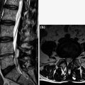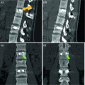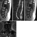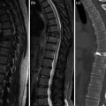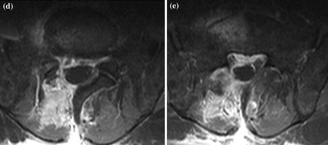
Fig. 1
a–e. SE T1 (a) and fat sat FSE T2 (b) sagittal, CE fat sat SE T1 sagittal (c) and axial (d–e). Right L5-S1 dural sac compression due to material adjacent to the disk and with its same MR signal. c–d: recurrent hernia surrounded by CE granulation tissue with involvement of the rear surgical breach. Both nerve roots at lower level are cleary visualized (e)
< div class='tao-gold-member'>
Only gold members can continue reading. Log In or Register to continue
Stay updated, free articles. Join our Telegram channel

Full access? Get Clinical Tree


