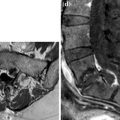
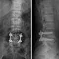
Fig. 1
a–b. Lumbosacral XR: posteroanterior (a) and lateral (b) views. L5–S1 diskectomy, intervertebral cage, and L4-S1 posterior stabilization
Postoperative Follow-Up After 6 Months


Fig. 2
a–b. Lumbosacral dynamic XR: lateral views in hyperflexion (a) and in hyperextension (b). No evidence of instability, preserved alignment of posterior vertebral profile
Postoperative Follow-Up After 9 months


Fig. 3
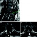
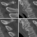
a–m. MPR sagittal (a–e) and axial sections at L4–L5 (f–i) and L5-S1 (j–m). Post-surgery inhomogeneity of retrovertebral space due to the presence of fibrotic tissue (a–e). Axial images at L4–L5 (f–i) document angulation of right pin with respect to its screw (arrow); axial images at L5-S1 (j–m) document the cause of low back pain due to left S1 root surrounded by fibrous scar (arrowheads)
< div class='tao-gold-member'>
Only gold members can continue reading. Log In or Register to continue
Stay updated, free articles. Join our Telegram channel

Full access? Get Clinical Tree


