Herniation
Subfalcine herniation is the most common brain herniation, and is caused by a supratentorial mass on one side that shifts brain to the opposite side. Herniation of brain occurs across the midline under the inferior or “free” margin of the falx. The cingulate gyrus, anterior cerebral artery, and internal cerebral vein can be displaced to the contralateral side under the falx. The ipsilateral ventricle will be compressed and displaced toward the midline. The contralateral ventricle can enlarge (unilateral obstructive hydrocephalus), due to occlusion of the foramen of Monro. The herniated anterior cerebral artery can also be compressed against the free margin of the falx, and thus occluded with subsequent infarction of the cingulate gyrus.
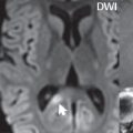
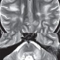
Descending transtentorial herniation is the second most common cerebral herniation, and can caused by a large hemispheric mass, occurring subsequent to subfalcine herniation. The uncus of the temporal lobe will be displaced medially and encroach upon the suprasellar cistern. On imaging, both the ipsilateral ambient cistern (which lies lateral to the midbrain) and lateral portion of the prepontine cistern may be widened. With further mass effect, the uncus and hippocampus can both herniate through the tentorial incisura. Descending transtentorial herniation can be either unilateral or, in more severe cases, bilateral ( Fig. 1.54 ).
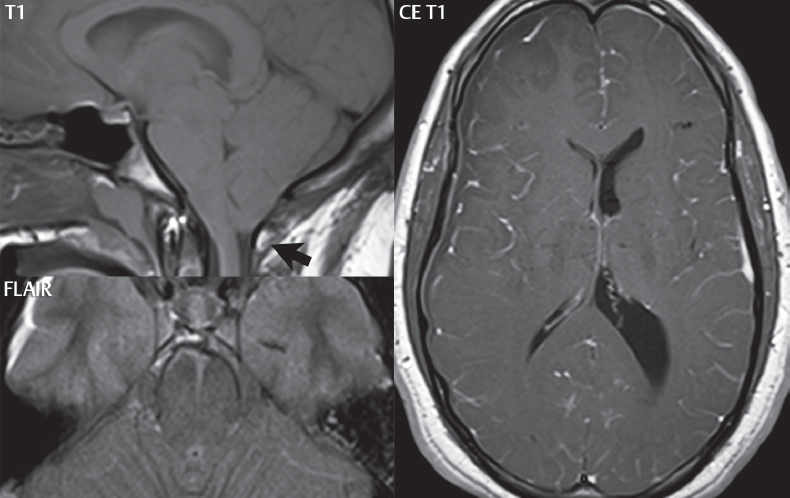
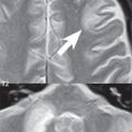
Tonsillar herniation is well visualized by MR, with the imaging findings including inferior displacement of the tonsils, pointing of the tonsils (in distinction to their normal rounded shape), and obliteration of CSF at the level of the foramen magnum ( Fig. 1.54 ).
Related posts:

Stay updated, free articles. Join our Telegram channel

Full access? Get Clinical Tree


