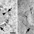
Fig. 1.
(a) shows an overview and (b) a higher magnification of two tomograms of an M2 macrophage 5 days after infection with the endotheliotropic HCMV strain TB40E at moi 5. Secondary envelopment of the viral capsids takes place in the assembly complex in the cytoplasm of an infected cell. The arrows depict the same viral particle in the three section planes. MTOC: microtubule organizing center. Section thickness is 570 nm as measured in the microscope.
3.5 Tomogram Reconstruction and Visualization
For tomogram reconstruction, we use the IMOD software (13). Tomograms are reconstructed by weighted back projection (WBP) or by simultaneous iterative reconstruction technique (SIRT) with 25 iterations using the standard settings of the IMOD software package version 4.1.2. For segmentation and data display, we use the AMIRA or the IMOD software.
4 Notes
The presented tomography method by tilting a semi thin section in the electron beam and reconstructing the data to a three-dimensional tomogram is currently challenged by block face methods. These methods work on the principle of classical SEM, imaging the trimmed blockface of a stained and resin embedded sample, and then gradually remove some resin before taking the next picture. Resin is removed either by focused ion beam (21) or by ultra thin sectioning using an ultra microtome in the SEM (22). These methods have the advantage that the process can be iterated many times and, therefore, much larger volumes can be analyzed than with STEM tomography, where section thickness is limited to about 1 μm, as explained above. The ultrastructural resolution of STEM tomography is, however, still considerably better, especially in X and Y direction.
Acknowledgment
We thank Li Wang, Ulm University for providing the sample shown in the figure.
References
1.
2.
Hoppe W et al (1974) Three-dimensional reconstruction of individual negatively stained yeast fatty-acid synthetase molecules from tilt series in the electron microscope. Z Physiol Chem 355:1483–1487
3.
Midgley PA, Weyland M, Thomas JM, Johnson BFG (2001) Z-Contrast tomography: a technique in three-dimensional nanostructural analysis based on Rutherford scattering. Chem Commun 10:907–908CrossRef
4.
5.
6.
Related posts:
 Histochemical Detection of Lipid Droplets in Cultured Cells
Environmental Scanning Electron Microscopy Gold Immunolabeling in Cell Biology
Histochemical Detection of Lipid Droplets in Cultured Cells
Environmental Scanning Electron Microscopy Gold Immunolabeling in Cell Biology
 Colocalization Analysis in Fluorescence Microscopy
 Morphological Analysis of Autophagy
 Environmental Scanning Electron Microscopy in Cell Biology
Stay updated, free articles. Join our Telegram channel

Full access? Get Clinical Tree


