, Joon Woo Lee1 and Eugene Lee2
(1)
Department of Radiology, Seoul National University College of Medicine, Seoul National University Bundang Hospital, Seongnam, South Korea
(2)
Department of Radiology, Seoul National University Bundang Hospital, Seongnam, South Korea
It is important to understand the histologic basis for the imaging appearances of spinal tumors in order to arrive at the correct diagnosis or formulate reasonable differential diagnoses. For example, hemangiomas in the vertebral bodies commonly contain fatty stroma and reveal high T1 and T2 signal on MR imaging, which is the main clue for its diagnosis. Highly cellular tumors such as lymphomas show intermediate signal intensity on T2-weighed images, which is also a clue for its diagnosis.
2.1 Tumors with Fatty Component
Fatty components within tumors show high signal on T1-weighted and T2-weighted MR images and can be suppressed with fat suppression MR techniques. The most common tumor containing fatty component is a hemangioma. If we see a fat-containing tumor in the vertebral body, the most probable diagnosis is that of a hemangioma. However, note that in contrast to vertebral hemangiomas, epidural hemangiomas do not show fatty signal in most cases. The most common fat-containing tumor in the epidural space is an angiolipoma, while the most common fat-containing tumors in the paravertebral muscles are lipomas and liposarcomas.
2.2 Red Marrow Component
Red marrow hyperplasia can mimic bony metastasis. Red marrow can show intermediate signal on T1-weighted images with patchy areas of slight enhancement. The signal of red marrow should be isointense or hyperintense to that of the intervertebral disc on T1-weighted images. Similar signal can be seen in multiple myeloma; however, multiple myelomas show strong enhancement in most cases.
2.3 Tumors with Vascular Component
Vascular components of spinal tumors show strong enhancement similar to those seen in venous structures on MRI. Usually, these vascular components show high signal intensity on T2-weighted images; however, in cases of intratumoral hemorrhage areas of low signal intensity can be seen on T2-weighted images. Common vascular tumors of the bony vertebrae include hemangiomas, telangiectatic osteosarcomas, and aneurysmal bone cysts. The common vascular tumors in the epidural space are hemangiomas and angiolipomas. The most common vascular tumor in the intradural extramedullary space is a paraganglioma, and the most common vascular tumor in the spinal cord is a hemangioblastoma.
2.4 Tumors with High Cellularity
Highly cellular components of tumors show intermediate signal intensity on T2-weighted images and demonstrate enhancement. This is often seen in lymphomas and meningiomas and can be found in some highly cellular metastases or sarcomas.
2.5 Tumors with Hemorrhagic Component
Hemorrhagic components within tumors can show variable signal intensity depending on the stage of bleeding. Chronic repetitive bleeding can result in hemosiderin deposition within or around the tumor. Hemosiderin is of dark signal on T1-weighted and T2-weighted images. Such signal change can be exaggerated on gradient echo images due to susceptibility blooming artifacts.
2.6 Tumors with Calcification/Ossification
Calcification/ossification can be more easily and reliably detected on CT compared to MR images, demonstrating high attenuation on CT images while on MRI showing low signal intensities on all sequences. Intratumoral calcifications can be seen in chondrosarcomas and meningiomas, while intratumoral ossification can be found in osteoblastomas and osteosarcomas.
2.7 Illustrations: Histologic Basis for Imaging Appearances of Spinal Tumors
2.7.1 Tumors with Fatty Component

Fig. 2.1


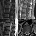
Hemangioma at L1 vertebra in a 57-year-old woman. Axial CT scan of lumbar spine (a) shows an osteolytic lesion in the left posterior corner of the vertebral body with internal dot-like trabeculation (white arrows). T1-weighted sagittal (b) and T2-weighted sagittal (c) MR images show high signal intensities indicating internal fat component with preserved coarse trabeculation (black arrows). Contrast-enhanced T1-weighted sagittal MR image (d) shows avid enhancement
Stay updated, free articles. Join our Telegram channel

Full access? Get Clinical Tree


