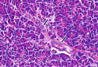
Fig. 3.1
This histologic section from the tail of the pancreas highlights the lobular architecture of the pancreatic parenchyma. Several acini (exocrine) drain into small interlobular ducts (short arrows), which drain into a large duct (long arrow). The endocrine portion is characterized by several islets of Langerhans (arrowhead) (H&E, 2.5×)
The predominant component of the exocrine portion are the acini.
The islets of Langerhans compose the endocrine portion
3.2.1 Exocrine Pancreas (Figs. 3.1–3.5)
Acini
Multiple acinar cells form the acini, which are the secretory units of the exocrine pancreas.
Each acinus consists of a single layer of acinar cells organized around a central lumen.
Acinar cells are polarized epithelial cells with a well-developed rough endoplasmic reticulum and cytoplasmic secretory granules called zymogen granules (Fig. 3.2).
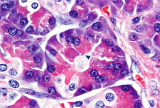
Fig. 3.2
Several acinar cells characterized by a pyramidal shape with round central nucleus, basophilic cytoplasm at the base, and eosinophilic granules (arrowhead) toward the lumen forming an acinus. These granules are rich in zymogens such as trypsinogen, chymotrypsinogen, procarboxypeptidase, proelastase, lipase, amylase, prophospholipase, and kallikreinogen (H&E, 100×)
These secretory granules are located in the apical part of the cell and contain multiple enzymes required for digestion.
Acini make approximately 85 % of the mass of the pancreas.
Ductal system
The ductal system begins within the central lumen of the acinus that connects to an intercalated duct (Fig. 3.3).
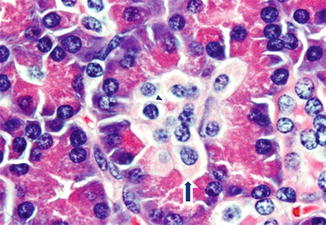
Fig. 3.3
Multiple small cuboidal cells in the center of the acinus with a pale glycogen-rich cytoplasm (arrow) converge to form an intercalated duct (arrowhead) are shown. These cells are in charge of chloride, bicarbonate, and water secretion to buffer and stabilize the zymogens until activation in the duodenum takes place (H&E, 100×)
Each intercalated duct consists of a single layer of centroacinar cells.
Centroacinar cells are cuboidal cells with few organelles and a pale cytoplasm.
Each pancreatic lobule is drained by the fusion of multiple intercalated ducts into a larger intralobular duct (Fig. 3.4).