
Historical Perspective of the Components of Image-Guided Neurosurgery
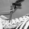
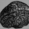
It is an essential aspect of neurosurgery that every cranial procedure must be conducted with the utmost precision. The brain, as opposed to any other organ in the body, is highly organized into structures that serve specific and irreplaceable functions. Collateral damage to these structures in the surgical pursuit of a deep target carries obligatory neurological deficits that are often permanent, and inexcusable. Further the external appearance of the brain gives little in the way of clues to the function subserved within, providing neurosurgeons with a formidable challenge to achieve intraoperative localization. The duration of training of every neurosurgeon is testimony to the challenge of achieving a sense of precise cranial navigation that is based in a firm comprehension of the three-dimensional (3-D) organization of the human brain. However even a perfect and encyclopedic knowledge of the normal functional anatomy of the brain is inadequate when confronted by the need to operate upon a brain altered either through normal variation or the presence of a disease process. Given the ability of the brain to compress, shift, and distort in the presence of disease it follows that neurosurgeons rely upon preoperative imaging more than any other surgical subspecialty in preparation for a procedure upon the brain to contort their concept of normal anatomy to match the distorted anatomy of the patient.
Neurosurgeons have been the beneficiaries of a rich variety of choices in the imaging of the brain. Computed tomography (CT), followed by magnetic resonance imaging (MRI), has most recently provided detailed 3-D images to guide cranial procedures. Even before the advent of these inherently three-dimensional techniques, neurosurgeons developed and were the first to use contrast techniques that improved the ability of two-dimensional radiographs to depict the position of the brain. Dandy’s use of pneumoencephalography, and Muniz’s development of cerebral angiography serve as testimony to the dependence of even veteran and experienced neurosurgeons to alter their well-developed visualization of the brain to match the actuality of a specific patient. In a very real sense neurosurgeons have always sought, and even striven, to be guided by images as they take on the challenges of operating on the brain. Indeed the apparent novelty of the concept of image guidance is a non sequitur, in that all surgery is image guided as surgeons operate guided by images of the surgical field formed on their retinas. It would therefore appear to be presumptuous to suggest that the history of image guidance is somehow distinct from the history of neurosurgery itself, as the two are essentially inseparable and likely synonymous. However, as recent developments in diagnostic imaging offer the potential of extending the vision of the surgeon far beyond the surface of the surgical field, the true power of image guidance will become increasingly apparent.
If a distinction can be made between the histories of image guidance and neurosurgery it can be found within the technology underlying image guidance, and the individuals who adapted said technology to fit the environment of an operating room. Given the challenges of neurosurgical procedures, it is to be expected that neurosurgeons, in addition to the ability to think in three dimensions, have strong personalities. Only individuals confident in their own abilities could enter the brain, address a problem within this most complex and least understood structure known in the universe, and tolerate horrific and disabling complications that would strip less confident individuals of any desire to take on such a challenge again. Given the confidence and self-reliance integral to their personalities it is common for many veteran neurosurgeons to dismiss intraoperative technological advances as being the unreliable infrastructure required by less confident, and perhaps less talented, surgeons to bolster their attempts to perform cranial surgery. Just as the microscope was seen as an unneeded operative toy when it was first employed intraoperatively, the proponents of image guidance and its predecessor stereotactic surgery were ostracized from the mainstream of neurosurgical history. This phenomenon was perhaps most notable in Europe, where entire departments of stereotactic surgery were founded outside existing departments within neurosurgery. Therefore, if image guidance has a history separate from that of neurosurgery, it is one created by the enmity of the majority of the neurosurgical community toward individuals who, rather than being uncertain of their surgical skills were beholden to the tremendous responsibility of operating on the seat of a patient’s psyche.
The history of image guidance consists of the stories of individuals confident not only in their surgical capabilities but in their ability to apply technological advances to reliably assist in navigation through the brain. These individuals could see beyond the limitations of existing techniques and apply untested technology to remove these limitations. As a result, neurosurgical procedures became more complex as new techniques and instrumentation supplanted preexisting technology. This increased complexity of cranial procedures, albeit a drawback, diminishes the discomfort and complications of conventional surgery and therefore is justified by the improved outcome for patients. Indeed, the majority of neurosurgeons have adopted these techniques. Just as any contemporary neurosurgeon would reject replacement of modern and complex powered drills by the simple trephines of yesteryear, so will the neurosurgeon of the future refuse to operate without the presence of an operative computer. (As all recent advances of new imaging technologies are based on the use of computers, the computer has become a new instrument in the operating room. Thus, an alternative name for the field could be computer-assisted surgery, although this appellation stresses the means over the benefits of this technique.) As minimally invasive procedures are more broadly adopted, there will be less opportunity for conventional orientation during surgery and a greater reliance on guiding technology and its inherent complexity.
This chapter defines the processes unique to image guidance—periprocedural information acquisition, information registration, and intraprocedural tracking—and uses this structure to recount the history of their development and provide a brief indication of the probable future of each process.
 Periprocedural Information Acquisition
The name image-guided surgery implies the coupling of some form of imaging to the surgical act and is the first essential component of such procedures. The history of image guidance is therefore inherently intertwined in the history of radiology as successive imaging advances underwent modifications to allow their use either prior to or during surgery. A history of radiology is beyond the scope of this chapter; but these modifications of imaging technology are central to the history of the topic.
It is important to note that the term image-guided surgery is at best a misnomer. Surgeons are not necessarily guided by images themselves, but rather by the information contained therein. Therefore, a more appropriate term for the field would be information-guided therapy, acknowledging not only the role of information but also the fact that many therapies that would benefit from such a coupling are not surgical in nature. Because these techniques can be used broadly with any invasive therapy that produces local effects with minimal collateral damage, newer forms of radiation therapy, such as particle beam therapy, can benefit by the use of identical visualization techniques. Further, the nature of the information employed can be quite diverse. The customary means of acquiring information is to subject the patient to a process that extracts the information from the anatomy of the patient. Usually the information extracted is organized in a 3-D virtual structure matched as precisely as possible to the anatomy of the patient, such as a CT or MRI scan. In other less demanding situations it is sufficient to employ two-dimensional information obtained by projecting the anatomy of the patient onto a single plane. In either situation, the resultant information can be categorized as an information data set, emphasizing the fact that the extraction process renders data that are an incomplete representation of the reality of the patient. The fidelity of the extraction process largely determines the utility of the resultant information to guide therapy. Given these considerations the term information-guided therapy will be used throughout the rest of this chapter to describe this field.
An important alternative to the extraction of data from the patient is to match the patient to a preexisting source of information obtained through the study of prior individuals. This preexisting repository of information is usually organized three-dimensionally and is termed an atlas. Atlas-based interventions suffer inaccuracy due to improper matching of the patient to the atlas or to inherent variability between individuals that diminishes the resolution of the atlas-based information. As our ability to extract information from patients improves, it can be anticipated that atlas-based interventions will be replaced; but in the early history of the field, atlases were the only source of information available. Because atlas-based interventions do not employ any data extraction from the patient, the history of the field actually predates the invention of imaging as we currently use the term.
The history of atlas-based interventions starts in 1889 with Dr. Zernov, a Russian surgeon who proposed the first device that superimposed a coordinate system on the patient’s head for localization of brain structures.1 It is of great interest that the information employed consisted of a phrenological atlas relating the contour of the patient’s head to prior patients with known psychological conditions. Dr. Zernov hoped to employ this matching process to guide the production of lesions within the brain to resolve the psychological condition implied by this matching process. Although his confidence in the technology did not allow him to employ the device clinically, his use of contour matching was the first instance of a registration technique that is enjoying a resurgence of interest in contemporary systems. In 1873, another early investigator, Dr. Dittmar, created the first device designed to develop a stereotactic atlas.2 He proposed investigating the function of the spinal cord in the rat by using his apparatus to insert a blade into the structure and recording the subsequent neurological deficit. Once again it was hoped that by matching the patient’s anatomy to the resultant atlas, information about the deficit experienced by the patient could be obtained, with essentially no concept of imaging being employed in the process. Atlas-based technology reached its zenith after 1952 when Spiegel and Wycis published the first stereotactic brain atlas for operative use.3 Subsequently numerous other stereotactic brain atlases have been produced since the early days of stereotactic neurosurgery. This topic is reviewed in Chapter 19 of this book.
Related posts:

Stay updated, free articles. Join our Telegram channel

Full access? Get Clinical Tree


