KEY FACTS
Terminology
- •
Congenital anomaly in which kidneys are fused at their lower poles in midline
Imaging
- •
Kidneys on opposite sides with lower poles fused in midline
- ○
Note that in crossed-fused ectopia, both kidneys are located on one side
- ○
- •
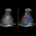
Presence of isthmus is defining feature; isthmus may be fibrous or functioning renal tissue
- ○
Isthmus crosses midline anterior to spine and aorta but posterior to inferior mesenteric artery
- ○
- •
Cross-sectional imaging (CT or MR) to define vascular anatomy and can be used to visualize associated complications such as malignancy
- •
May be first detected by ultrasound but vascular anatomy better defined by CTA or MRA
- •
Arterial supply and venous drainage may be complex
Top Differential Diagnoses
- •
Crossed-fused ectopia
- •
Renal ectopia
- •
Paraaortic lymphadenopathy/retroperitoneal mass
Pathology
- •
Fusion of kidney precursors in early gestation when still located in pelvis, preventing appropriate ascent
Clinical Issues
- •
1 in 500 children; M:F = 2:1
- •
30% asymptomatic
- •
Up to 2/3 have associated abnormalities (most commonly genitourinary)
- •
Complications include uretero-pelvic junction obstruction, infection, nephrolithiasis
- •
Increased risk of malignancy such as Wilms tumor, urothelial carcinoma, and carcinoid
- •
Prognosis is good in absence of associated abnormalities
Scanning Tips
- •
Suspect when lower renal poles disappear medially
- •
Look for isthmus in midline anterior to aorta and posterior to inferior mesenteric artery
- •
Use compression to displace bowel
 anterior to the aorta and inferior vena cava. Note the additional renal arteries arising from the common iliac arteries
  .
.
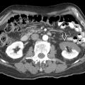
 of the horseshoe kidney lies anterior to the spine
  and vena cava
  . The isthmus is composed of functioning renal tissue.
 of the horseshoe kidney lies anterior to the aorta
  and inferior vena cava
  . The isthmus is composed of functioning renal tissue.
Stay updated, free articles. Join our Telegram channel

Full access? Get Clinical Tree


