CT
Rectum (Dose in cGy/fx)
Bladder (Dose in cGy/fx)
Femoral heads (Dose in cGy/fx)
Diagnostic multidetector CT
1.74
1.83
4.05
kV CBCT
1.70
1.73
3.14
MVCT
1.05
1.04
1.02
2 Clinical Implications
Because of the relatively new employment of the previously described IGRT techniques in the daily routine, there is a lack of data regarding the clinical implications and reduction of toxicities in comparison with non-IGRT approaches. Thus far, studies have shown that these strategies are feasible in terms of treatment accuracy, with the attendant reduction in margins and radiation exposure to nearby critical organs.
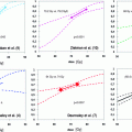
Table 2 depicts the mean CTV to PTV margins when different daily IGRT techniques are employed (Beltran et al. 2008; Johnston et al. 2008; Gayou and Miften 2008).
However, whether these improvements lead to reduced toxicities, as reported by patients, is uncertain. Song et al. performed an evaluation of image-guided radiation therapy technologies and their impact on the outcomes of hypofractionated prostate cancer treatments by using NTCP and TCP models (Song et al. 2006). The tattoo registered technique had a significant reduction in TCP as the prescription dose decreased. The differences between the techniques other than the tattoo registered (image-guided alignment to bony landmarks, to the daily CTV volume, or alignment to the CTV volume with daily monitor units updates), however, were small (<2.7 %) (Song et al. 2006).
No randomized trials are available. For the existing data, greater patient populations and longer follow-up is needed to determine the importance of IGRT in reducing the acute and chronic toxicities. A study by Jereczek-Fossa et al. (Jereczek-Fossa et al. 2011) assessed in a nonrandomized fashion the acute toxicity of image-guided hypofractionated radiotherapy. 179 patients were treated within the prospective study with 70.2 Gy/26 fractions (equivalent to 84 Gy/42 fractions, α/β 1.5 Gy) using IGRT (transabdominal ultrasound, ExacTrac X-Ray system, or cone-beam computer tomography) techniques. The data of these patients were compared to retrospective data of non-IGRT patients. The acute toxicity rates were low and similar in both study groups (Jereczek-Fossa et al. 2011). Similar results were achieved in Klinkum rechts der Isar, Munich Germany (Geier et al. 2012).
3 Conclusion
An ideal IGRT system would allow for daily prostate imaging without possible introduction of errors due to image acquisition itself, it would do so within a reasonable time frame, without the necessity for implanted radio-opaque markers and preferentially without exposing the patient to radiation (Soete et al. 2008).
A solution that combines all these features is inexistent so far. For the existing IGRT techniques, there is a considerable lack of data whether they lead to a reduced acute and chronic toxicity profile in comparison with the non-IGRT approach, or if they are associated with an improved local control. Nevertheless, given the increasingly higher doses and smaller treatment margins utilized, combined with the trend to hypofractionate radiation therapy, daily IGRT for prostate cancer has become a necessity as an accurate and precise way of delivering the intended dose to the PTV and the OARs. The problem of interfractional prostate movement and the possibility of setup errors are optimally accounted for.
References
Cheng CW, Wong J, Grimm L, Chow M, Uematsu M, Fung A (2003) Commissioning and clinical implementation of a sliding gantry CT scanner installed in an existing treatment room and early clinical experience for precise tumor localization. Am J Clin Oncol 26:e28–e36PubMed
Related posts:

Stay updated, free articles. Join our Telegram channel

Full access? Get Clinical Tree


