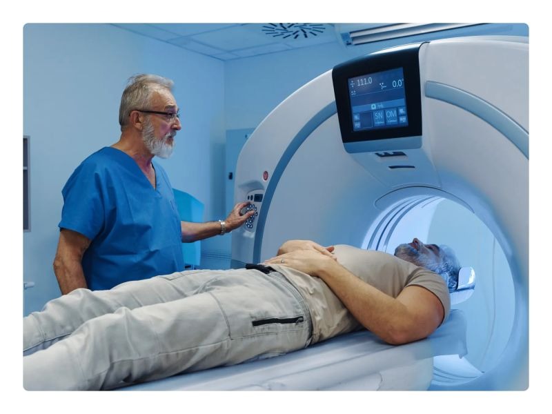
Emergency departments are busy, noisy, and unpredictable. Radiology sits at the center of many urgent decisions — stroke, trauma, chest pain, shortness of breath. Artificial intelligence (AI) is not a magic wand, but it is reshaping how images are moved, read, prioritized, and reported. The change is practical. It is also fast. Shorter waits. Fewer missed urgent findings. New kinds of mistakes. Let’s walk through what’s happening and why it matters.
What AI actually does in the ED (simple list)
- Flags urgent findings (e.g., intracranial hemorrhage, large pneumothorax).
- Prioritizes the worklist so a radiologist sees the most critical cases first.
- Pre-measures structures and creates structured data (volumes, scores).
- Automates routine tasks (protocol selection, image transfer, report drafting).
- Integrates clinical data with images to offer risk scores.
Short. Clear. Useful.
Triage and prioritization — faster attention to life-threatening problems
AI triage tools monitor incoming CTs and X-rays and push alerts when they detect likely critical findings. That means radiologists can spot an intracranial bleed or a tension pneumothorax earlier and act faster. Real-world deployments have shown meaningful drops in reporting turnaround time for ICH (intracranial hemorrhage) and trauma — sometimes cutting average reporting times by more than half and increasing the proportion of critical reports completed within guideline windows.
Automated image analysis — a second pair of eyes
Deep learning models detect patterns (nodules, consolidation, fractures) that can be hard to spot in a busy ED. For chest X-rays, multicenter evaluations show high sensitivity and specificity for urgent abnormalities when AI is used as an assistive tool. That doesn’t replace the radiologist; it augments them — reducing misses particularly when workload or fatigue is high.
Workflow automation and report drafting — reducing grind work
AI can populate measurements, pre-fill report templates, and even draft narrative text from structured findings. More advanced systems help pick the correct imaging protocol at the scanner (MRI/CT parameter selection), reducing rescans and delays. Hospitals that implemented end-to-end AI workflow tools report large drops in mean reporting times (for certain trauma and ED cohorts) and big improvements in meeting critical-case time targets.
Adoption and scale — how common is this now?
Adoption is growing quickly, but it varies by region and hospital type. Recent surveys of radiologists and hospital reporting show a marked rise in clinical use compared to a few years ago, and national data indicate increasing uptake of predictive and diagnostic AI across hospitals. In short: some centers use many AI tools every day; others are still piloting a first model.
Concrete benefits (numbers you can use)
• Faster turnaround: studies and audits have recorded reductions in median reporting times from around an hour to under thirty minutes for prioritized ED cases after AI triage was switched on.
• Higher detection in certain settings: AI-assisted readings for urgent chest X-ray findings have reported sensitivities in the 90%+ range in some real-world datasets.
• Wider system gains: automating protocol selection and reporting tasks can free technologist and radiologist time, which can be redeployed to complex cases or direct patient care. (Numbers differ by site and the exact tools used.)
Workflow changes you’ll notice on the floor
- The worklist will reorder itself (sometimes every few minutes).
- You’ll get push alerts when a scan looks critical.
- Prepopulated reports shorten dictation time.
- Radiographers may receive AI-guided prompts at the scanner.
Small changes, collectively large in impact.
Security, privacy and communication concerns
AI tools require image access and many ties into electronic health records. This raises data-governance work: access controls, auditing, and vendor contracts. Also—and this is important for any staff discussing cases online—avoid sharing identifiable clinical images or patient data over unsecured channels.
If you ever discuss cases in informal online spaces, do so safely; consider privacy tools or apps for anonymous group chats to avoid any identifiable content. Anonymous online chats aren’t about sharing visitors’ personal information, but about sharing experiences without harming patients. Using privacy-focused video chats like CallMeChat is simply a secure channel for connecting with people. You never know who you’ll encounter next, but the experience promises to be engaging.
Human and technical risks (be blunt)
AI is fallible. False positives can create “alert fatigue” and slow things down. False negatives — missed findings — are the worst-case clinical risk. Regulatory status, validation, local calibration, and clinical governance matter a lot. Legal responsibility usually still rests with clinicians, not the algorithm. Explainability is limited for some models, which raises trust issues when a discordant case occurs.
Training, acceptance and workflow redesign
Adoption is not plug-and-play. Radiologists and ED teams need training, new protocols, and agreed escalation paths for AI alerts. That means workflow redesign: who responds to an AI “red flag”? How are false positives handled? What auditing loop is in place? Without these, tools underperform.
What’s next?
Expect more multimodal tools that combine images, labs, and notes to generate a risk estimate. Expect better integration with ED dashboards so bed managers and consultants see imaging priorities in real time. And expect ongoing studies that measure clinical outcomes — not just speed: does AI use actually improve morbidity and mortality for stroke, trauma, sepsis? Early evidence is promising but not yet definitive across all conditions.
Practical takeaways (quick)
- AI helps prioritize. Big wins there.
- AI helps with routine measurements and report drafting. Time saved.
- AI is not autonomous in most ED settings; human oversight remains essential.
- Validate and govern every tool locally. Don’t assume external validation equals local performance.
Conclusion
Artificial intelligence is changing emergency-department radiology in measurable ways: faster prioritization, shorter reporting times for critical cases, and automation of repetitive tasks. The technology amplifies human capability when properly integrated and governed. But it also introduces new failure modes and governance needs. The future is collaboration: smart algorithms + trained clinicians = safer, faster imaging care in the ED.
Related posts:
 Other Imaging Techniques in Dilated Cardiomyopathy
 Basic Echocardiography in Arrhythmogenic Right Ventricular Cardiomyopathy
 Early Diagnosis of Sports Injuries: Why Timely Imaging Matters
Enhancing Diagnostic Accuracy Through Online Radiology Consultation on Ai PACS
Dental Imaging Explained: From Bitewings To Panoramic X-Rays
Support Systems That Help Working Nurses Succeed in School
Early Diagnosis of Sports Injuries: Why Timely Imaging Matters
Enhancing Diagnostic Accuracy Through Online Radiology Consultation on Ai PACS
Dental Imaging Explained: From Bitewings To Panoramic X-Rays
Support Systems That Help Working Nurses Succeed in School
Stay updated, free articles. Join our Telegram channel

Full access? Get Clinical Tree


