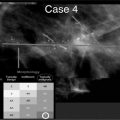
ACR grading of mammographic breast tissue density
Glandular tissue component (%)
Description
I
<25
Almost entirely fat
II
25–50
Scattered fibroglandular densities
III
51–75
Heterogenously dense
IV
>75
Extremely dense
Perception is also influenced by the tumor growth pattern. The most frequently occurring histological types of breast cancers are invasive ductal and intraductal cancers in about 75% of patients, followed by invasive lobular cancers in 5–15%, mucous and medullary cancers in 5– 7%, tubular cancers in 2–6%, and papillary cancers in about 2%. The cancer growth pattern may frequently produce an irregular-shaped mass with spiculations and microlobulations (“hedgehog type”), a more lobulated “benign looking” circumscribed mass (“potato type”), and several types of masses created by a mix of “hedgehog” and “potato”. In addition, two different growth patterns can be described: the intracystic growing cancer and the diffuse growth type, a mix of two or more different growth patterns can exist in one cancer. The most challenging cancer growth pattern to perceive in mammograms is the diffuse type. This type has the tendency to produce nonmass asymmetries that are sometimes visible in only one mammographic view, or it can result in architectural distortion. Invasive lobular cancers can show a more diffuse growth pattern, with tissue stiffening. Frequently, additional physical examination shows harder asymmetric palpable breast tissue or an asymmetric palpable mass in the mammographic region of interest.
In population-based mammography screening programs, routine mammography double reading has been proven to increase the number of visible cancers and detected cancers by 5–15% compared with single reading. In addition, computer-aided detection (CAD) can be used to reduce the rate of missed cancers. In the literature, the sensitivity of CAD for masses has been reported to be in the range 67–89% [4–6]. Gilbert and colleagues performed a retrospective comparison of single reading with CAD and double reading without CAD [7]. They showed that single reading with CAD led to a significantly higher cancer detection rate than double reading alone. The single reading with CAD found 6.5% more cancers than the double reading alone. The negative finding was an increase in recall rate for single reading plus CAD (8.6%) compared with double reading (6.5%).
Assessment
The perturbations in a mammogram can be caused by calcifications, masses, focal or global asymmetries, architectural distortions with or without contour deformity, tubular densities or dilated ducts, nipple and/or skin retraction, skin thickening, skin lesions and axillary adenopathy. The ACR and the Breast Imaging Reporting and Data System (BI-RADS®) [2] have standardized the assessment of lesions found by mammography, ultrasound and magnetic resonance imaging (MRI) in the BI-RADS® lexicon. In the next chapter, the mammographic BI-RADS® assessment terminology of masses, architectural distortion and asymmetries will be highlighted. Table 2 presents the BIRADS ® descriptors and BI-RADS® terminology (valid fourth edition terminology) concerning mass and architectural distortion. At the meeting of the Radiological Society of North America (RSNA) 2012, Sickles presented the changes that are expected in the fifth edition of the BIRADS ® lexicon, which is planned for publication in 2013. These expected changes concerning the BI-RADS® desciptor “asimmetry” are implemented.
Table 2
Mammography assessment of mass and architectural distortion using BI-RADS® descriptors (fourth edition terminology)
BI-RADS® descriptor | ||
|---|---|---|
Mass | Shape | Round |
Oval | ||
Lobular | ||
Irregular | ||
Margins | Circumscribed | |
Indistinct or ill defined | ||
Microlobulated | ||
Spiculated | ||
Density High | Equal | |
Low | ||
Fat containing | ||
Architectural distortion | Normal breast architecture disturbed | |
Special cases | Intramammary lymph node | |
Tubular density or dilated duct | ||
Global asymmetry | ||
Focal asymmetry | ||
Associated findings | Skin retraction | |
Nipple retraction | ||
Skin thickening | ||
Trabecular thickening | ||
Skin lesion | ||
Axillary adenopathy | ||
Masses
According to the BI-RADS® Breast Imaging lexicon, a mass is defined as a three-dimensional (3D) structure with convex outward borders. Typically, a mass can be visualized on two orthogonal views. A mass with round or oval shape combined with a circumscribed margin and low or equal density (compared with breast parenchyma) has a high probability of being benign. A high-density mass with irregular shape and spiculated margin is suspicious for breast cancer. To call a margin “circumscribed”, more than 75% of the mass has to be free from superpositions. Lazarus and colleagues studied the interobserver variability of different mammographic masses related BIRADS ® descriptors [8]. A moderate agreement was obtained for mass shape (κ=0.48) and mass margin (κ=0.48), and a slight agreement for mass density (κ=0.18). Concerning the fact, that a mass is perceived by the readers, a high κ-value of 0.84 resulted. Masses with indistinct margins can start initially as asymmetries.
Stay updated, free articles. Join our Telegram channel

Full access? Get Clinical Tree


