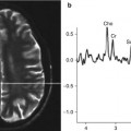
Fig. 4.1
Map of Europe. The status of central nervous system hydatidosis is discussed in the Eastern and Central European countries (sprayed on map) where data were available
Epidemiological and Clinical Features Regarding Hydatidosis of the Central Nervous System in Central and Eastern European Countries
Hydatidosis in Romania
Epidemiology
In 1995, Romania was in the forefront of the European countries and among the first countries worldwide as regards the incidence of hydatid disease (Neghina et al. 2010, 2011a). Moreover, the severity of hydatidosis in Romania is emphasized by the fact that at least one person from 45.5 % of Romanian localities underwent surgery for this condition (Iacobiciu et al. 2005; Neghina et al. 2010, 2011b). Following the analysis of the available global data, Stefanoiu concluded in 2000 that the number of human cases of hydatidosis in Romania surpassed the number of cases from Lithuania, Belarus, Poland, Slovakia, Ukraine, Bulgaria, Czech Republic, and Latvia combined (Iacobiciu et al. 2005; Neghina et al. 2010). Nevertheless, hydatidosis is not a notifiable disease in Romania, and no cases were mentioned in the annals of the World Health Organization (WHO) during the period from 1996 to 2000 (Iacobiciu et al. 2005; Neghina et al. 2010). Studies performed in the entire country have shown that the incidence of the disease decreased from 5.6 cases per 100,000 inhabitants during the period 1953 to 1963 to 2.6 cases per 100,000 inhabitants from 1987 to 1991 (Neghina et al. 2010, 2011b; Calma et al. 2011). The most recent studies undertaken in four counties of western Romania indicated an average incidence between 2.4 and 4.4 cases per 100,000 inhabitants (Calma et al. 2011, 2012; Moldovan et al. 2012).
Hydatidosis of the Brain and Cranium
In 1900, Thoma Ionescu, considered the godfather of Romanian surgery, presented at the International Congress of Medicine and Surgery held in Paris a case of cerebral hydatidosis that he successfully operated on through temporal craniotomy, and at the same time, he emphasized the possible surgical risks. Two years later, he published in a reputable national journal new and useful data on the topic of cerebral hydatidosis with particular emphasis on the case he had previously managed (Arseni et al. 1988). In 1905, Antoniu reported for the first time in Romania a rare case of cranial hydatidosis. At that time, only seven such cases were registered in the medical literature worldwide. His paper, entitled “Hydatid Cyst of the Cranial Bones,” represented a valuable starting point toward the knowledge of this pathology in Romania. The patient, hospitalized in Turnu Severin (southwestern Romania), presented with a single tumor that caused cerebral compression with epileptic crisis. Unfortunately, the patient died before the onset of any exterior signs suggesting cranial involvement (Arseni et al. 1988).
During the period of 1935–1968, there were 46 cases of cerebral hydatidosis reported in Romania (Arseni and Samitca 1957; Arseni et al. 1981) (Table 4.1). Information that is more detailed was available for the cases managed during 1935–1955 in a Neurosurgery Clinic in Bucharest. Of 2,226 cases with intracranial space-occupying disorders, 36 patients (1.6 %) were found with cranial and intracranial hydatidosis (Arseni and Samitca 1957). The most significant epidemiological and clinical characteristics of this study group are summarized in Table 4.2.
Table 4.1
Cases of cerebral hydatidosis in Romania during the period 1935–1968
Year(s) | Authors | Hospital, city | No. of cases | Specific comments | References |
|---|---|---|---|---|---|
1935–1955 | Arseni and Samitica | Neurosurgery Clinic, Bucharest | 36 | Presented in Table 4.2 | |
1954a | Marcovici and Steriade | Unspecified | 1 | Giant cerebral cyst, recidivated, with abscess | Arseni et al. (1981) |
1957a | Russu | Unspecified | 2 | Study in children. The prevalence of cerebral hydatid cysts was 5.4 % of all locations | Arseni et al. (1981) |
One of the cysts (very voluminous) developed in a child who previously underwent surgery for a hepatic hydatid cyst | |||||
1960a | Waitsuk, Andrasofski, and Mera | Neurosurgery Clinic, Targu Mures | 6 | One of the cysts was situated in the right premotor cortex and had calcified walls | Arseni et al. (1981) |
1968a | Theodorescu and Mitroi | Neurosurgery Clinic, Constanta | 1 | The cyst, about the size of a small fist, was located in the left parieto-occipital cortex | Arseni et al. (1981) |
At hospital admission, the patient was comatose with respiratory failure. Patient’s condition worsened as a consequence of a lumbar puncture performed 1 month later | |||||
The patient died | |||||
Total cases | 46 | ||||
Table 4.2
Main epidemiological and clinical characteristics of patients with cranial and intracranial hydatidosis managed during 1935–1955 in a Neurosurgery Clinic in Bucharest (Arseni and Samitca 1957)
Feature | Relevant statistics | Percent of total cases |
|---|---|---|
Gender | Male | 58 |
Age | <30 years | 73 |
Inhabitance area | Rural | 64 |
Duration of symptoms | <1 year | 78 |
Type of cyst | Unilocular | 81 |
Symptomatology onset | Insidious | 92 |
Symptoms | Headache | 59 |
Epilepsy | 50 | |
Outcome | Immediate postsurgical mortality | 9 |
Late mortality (>18 months) | 13 | |
Without neurological sequels (% of surviving patients) | 40 |
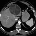
Until 1980, the number of cases with cerebral involvement rose to 83 in the Neurosurgery Clinic in Bucharest. Of these patients, 55.4 % were males and more than 43 % were children. The youngest patient was 1.5 years old, and the oldest one was 65 years old (Arseni et al. 1981). A further study in the same clinic presented the characteristics of 27 pediatric cases who underwent surgery during the period from 1980 to 1992 (Ciurea et al. 1995). An additional 52 cases diagnosed during the same period were not discussed due to different reasons. The studied cases represented 2.8 % of the children with expansive nontraumatic lesions hospitalized in this clinic. The mean age of the study group was 8.2 years, and the youngest patient was 3.3 years old. A 67 % male prevalence was registered, and most of the patients (41 %) belonged to the age group 6–10 years. The most prevalent symptoms were headache and vomiting (96 %), followed by papillary edema (89 %) and hemiparesis (70.3 %). In the majority of patients (40.7 %), two cerebral lobes were affected. Recurrences occurred in 40.7 % of patients (in all the cases where total removal of the cysts could not be achieved). Another study undertaken at the same clinic included 76 patients who underwent surgery for cerebral hydatidosis during the period from 1981 to 2003 (Ciurea et al. 2006a). The main epidemiological and clinical characteristics of the study group were the following: 95.7 % of patients were children; the median age was 8.7 years in children and 29.3 years in adults; male patients prevailed (59.2 %); increased intracranial pressure was the most frequent symptom in children (91.1 %), while headache was the most prevalent finding in adults (88.9 %); 94.8 % of the cysts were supratentorial; rupture of the cysts during the surgery was reported in 26.3 % of cases; postsurgical complications occurred in 67.1 % of patients; relapses developed in 25 % of cases; the outcome (either at 6 months or at 2 years) showed a fatality rate of 5.3 %.
Hydatidosis of the Spine and Spinal Cord
The first documented information regarding vertebro-medullary hydatidosis dates from the period 1862 to 1865, when Kalenderu reported two cases with vertebral cysts (detected in the dorsolumbar muscles) (quoted from Arseni et al. 1981). In 1898, Botescu communicated a case of hydatid cyst located in the pelvis, and Tzaicu reported in 1930 a case with medullary compression whose starting point was in the musculature and presented a secondary rachidian invasion (quoted from Arseni et al. 1981). In 1927, Jianu detected a case of vertebral hydatidosis and later a case of thoracolumbar involvement without neurological symptomatology (cases communicated in 1957) (quoted from Arseni et al. 1981). In 1946, the case of a patient with an epidural hydatid cyst was presented by Cosacescu and Vereanu. Later, in 1956, Horvath and Sandor presented their study on 14 patients with vertebro-medullary invasion (Arseni et al. 1981).
During the period from 1935 to 1957, a total of 20 cases of vertebro-medullary involvement due to hydatid cysts were noted in the Neurosurgery Clinic in Bucharest (Arseni et al. 1960). They account for approximately 3 % of cases with rachidian and intrarachidian tumors and approximately 0.06 % of all cases managed during this period. These cases represented about half the number of cases with cerebral hydatidosis. In most of the patients (40 %), the lesions were found in the lumbar region. The youngest patient was 14 years old and the oldest 66 years old. Male patients represented 55 % of the study group, and 80 % of the cases were rural inhabitants. It has been noticed that a high prevalence (25 %) of patients belonged to the 11- to 20-year-old age group. Radicular pain was the initial clinical symptom in most of the patients (70 %). However, syndrome of spinal cord compression was the most common finding (75 %) upon admission. The time span from the onset of the disease until hospital admission ranged between 0 and 6 months in the majority of the cases (40 %). Out of 19 cases who underwent surgery, the outcome was fatal in a single case; of the remaining patients who were operated and survived, relapses occurred in approximately 44 % of cases.
Throughout the time, other cases were also detected, as follows: a report on ten cases with bone involvement (of which four cases with vertebral involvement were diagnosed in the Department of Orthopedics of Brancovenesc Hospital, Bucharest) is presented by Negrea in 1957; in the same year, Vereanu reported four cases of bone involvement (including one case with vertebral location and one with sacral); in 1971, Marinescu reported 30 cases of vertebral hydatidosis (quoted from Arseni et al. 1981).
Craniospinal Hydatidosis
Lupascu and Panaitescu (1968) gave an overview in 1968, in their valuable treatise on hydatid disease, of the data reported by the National Cantacuzino Institute regarding involvement of different organs and systems in this disorder. According to this source, craniocerebral cases occurred with a prevalence of 1.4 %, and vertebral lesions were diagnosed in 0.7 % of patients.
A major epidemiological study performed in the entire country of Romania during the period from 1987 to 1991 (Iacobiciu et al. 2005), including 3,072 cases (2,701 adults and 371 children), provides valuable data on cerebral and vertebro-medullary involvement: 0.94 % of patients were diagnosed with the condition at first hospitalization, 0.16 % at the second admission, and 0.1 % at the third one (Table 4.3).
Table 4.3
Cerebral and vertebro-medullary involvement of hydatidosis in 3,072 patients hospitalized in all of the Romanian surgical sections between 1987 and 1991
Location of the hydatid cyst | Adults | Children | Total | |||
|---|---|---|---|---|---|---|
No. of cases | % of adult cases (n = 2,701) | No. of cases | % of children cases (n = 371) | No. of cases | % of total cases (n = 3,072) | |
First hospitalization | ||||||
Cerebral | 11 | 0.41 | 15 | 4.04 | 26 | 0.85 |
Hepatic and vertebro-medullary | 1 | 0.04 | – | – | 1 | 0.03 |
Thoracolumbar | 1 | 0.04 | – | – | 1 | 0.03 |
Only vertebro-medullary | 1 | 0.04 | – | – | 1 | 0.03 |
Total | 14 | 0.53 | 15 | 4.04 | 29 | 0.94 |
Relapse, second hospitalization | ||||||
Cerebral | 1 | 0.04 | 4 | 1.08 | 5 | 0.16 |
Relapse, third hospitalization | ||||||
Cerebral | – | – | 3 | 0.81 | 3 | 0.1 |
Further investigations in children up to 15 years old (407 patients, 1987–1992) indicated the following percentage of cerebral involvement according to age groups: 5.21 % (0–5 years), 5 % (6–10 years), and 1.8 % (11–15 years) (Iacobiciu et al. 2005). According to Gherman et al. (1991), the prevalence of cerebral hydatidosis was 1–2 % in 1991. In 1994, Stefanoiu and Vranceanu reported in their study a prevalence of 0.4 % for cerebral hydatidosis. In the same study, the prevalence of this condition was 4 % in 407 pediatric cases (Stefanoiu and Vranceanu 1994; Iacobiciu 1999). A survey taken from 1994 to 1999 in a southern Romanian county (Dolj county) reported cerebral involvement in 1 out of 213 cases (0.5 %) (Iacobiciu et al. 2005).
In 2000, Plesamosca and his collaborators reported data on 14 pediatric cases of hydatidosis with less common locations. Of these cases, cerebral involvement coexisted with involvement of other organs in 14.3 % of patients (Iacobiciu et al. 2005). A major epidemiological survey of 1,004 cases with hydatidosis diagnosed between 1985 and 1997 in three southwestern Romanian counties (Timis, Arad, and Caras-Severin) indicated cerebral involvement in 0.1 % of the study group (Iacobiciu 1999). The most recent studies undertaken from 2004 to 2010 in Timis (182 patients) (Calma et al. 2011), Arad (79 patients) (Calma et al. 2012), Caras-Severin, and Hunedoara counties (190 patients) (Moldovan et al. 2012) detected no cases with CNS involvement.
Hydatidosis in Former Yugoslavia and Countries Succeeding Former Yugoslavia
Yugoslavia
As reviewed in Arseni et al. (1981), in 1952, Stojanović and Vujadinović reported only a single case (2.3 %) with cerebral involvement out of 44 patients with hydatid disease (quoted from Arseni et al. 1981). Later, in 1957, Papo and Vrigisić found that of 200 cases in which patients underwent surgery, two patients (1 %) had hydatid cysts with cerebral location (quoted from Arseni et al. 1981). In the countries succeeding former Yugoslavia, data on cerebral echinococcosis are available for Serbia, Montenegro, Croatia, and Macedonia, whereas none are available for Slovenia and Bosnia and Herzegovina.
Serbia
Hydatidosis is a mandatory notifiable disease in Serbia, and consequently the relevant data on this topic are archived at the National Institute for Public Health (Tables 4.4 and 4.5). However, when comparing the official data to that published in the literature, it becomes obvious that cases are largely underreported to this institute. A clinical study on hepatic hydatidosis performed in the largest Serbian clinical center indicated that 1,016 adult patients underwent surgery for this illness between 1963 and 2006. Of this number of cases, 376 patients were managed between 1997 and 2006 (Basara 2007).
Table 4.4
Cases of hydatidosis registered in Serbia during the period 1991–2010
Year | Cases per 100,000 inhabitants | Number of cases | Gender distribution: male (%) |
|---|---|---|---|
1991 | 0.12 | 10 | NA |
1992 | 0.07 | 6 | NA |
1993 | 0.12 | 8 | NA |
1994 | 0.09 | 7 | NA |
1995 | 0.25 | 20 | NA |
1996 | 0.16 | 13 | NA |
1997 | 0.21 | 17 | NA |
1998 | 0.36 | 29 | NA |
1999 | 0.32 | 26 | NA |
2000 | 0.30 | 24 | NA |
2001 | 0.45 | 35 | 44.4 |
2002 | 0.36 | 27 | 31.4 |
2003 | 0.53 | 40 | 18.5 |
2004 | 0.28 |