Author(s), year
Country
Age and gender
Localization
Neurological status
Neuroimaging
Outcome
Bartel (1928)
France
NA
Cervical
NA
NA
NA
Deve (1928)
France
NA
Cervical, ID, EM
NA
NA
NA
Rauzier (1928)
France
NA
Cervical
NA
NA
NA
Vallè (1928)
France
NA
Cervico-thoraco-lumbar
NA
NA
NA
Bertrand et al. (1956)
Morocco
25, F
T11-T12 ID, EM
Paraplegia
Myelography
Recurrenceafter 13 mos
Boixados (1961)
Spain
4, F
T5 ID, EM
Paraplegia
Myelography
Partial recovery
Carrea and Murphy (1964)
Argentina
9, F
T4–T5 ID, EM
Paraplegia
Myelography
Total recovery
Pierini et al. (1965)
Argentina
NA
Cervical, ID
NA
Myelography
NA
Baurand et al. (1970)
North Africa
35, F
T6 ID, EM
Paraplegia
Myelography
Total recovery
Karvounis et al. (1977)
Greece
37, F
L5–S1 ID, EM
Sciatica
Myelography
Total recovery
Bettaieb et al. (1978)
Tunisia
4, M
Upper thoracic, ID, EM
Paraplegia
CT-myelography
Total recovery
Tunisia
8, M
Upper thoracic, ID, EM
Paraplegia
CT-myelography
Total recovery
Tunisia
8, M
Middle thoracic, ID, EM
Paraplegia
CT-myelography
Total recovery
San Martin Sánchez et al. (1980)
Spain
62, F
L2, ID, EM
Paraplegia
Myelography
Total recovery
Sharma et al. (1981)
India
14, M
T6–T10, ID, EM
Paraplegia
Myelography
Partial recovery
Pamir et al. (1984)
Turkey
34, F
L2, ID, EM
Paraplegia
Myelography
No change
Akhan et al. (1991)
Turkey
6, M
T9–T11, ID, EM
Paraplegia
CT-myelography
NA
Medjek et al. (1991)
Algeria
21, F
T12–L1, ID, EM
Paraplegia
CT-myelography
Total recovery
Fahl et al. (1994)
Haddad and Bitar (1997)a
Turgut (1997)b,c
Islekel et al. (1998)
Lebanon
55, F
T12–L1
Paraparesis
CT-myelography, MRI
NA
Lebanon
46, M
L4–S1
Sciatica
CT
Recovery
Lebanon
7, M, and 7, F
Cervical (2), thoracic (11),lumbar (4), sacral (1)
Pain (13), urinaryincontinence, weakness(5), sciatica (2)
Myelography (7),CT (4), MRI (2)
Death (2), totalrecovery (2),no change (1)
Turkey
61, M, and 23, F(mean age: NA)
Cervical (3), thoracic (35),thoracolumbar (3), lumbar(28), sacral (1), cervicaland lumbar (1)
Paraplegia and paraparesis (61), caudaequine syndrome (27)
Myelography (56),
MRI (8), CT (6),US (1)
Death (2), totalrecovery (5),recurrence (15)
Turkey
19, M
L2–L4, ID, EM
Paraplegia
Myelography
Partial recovery
Kabbaj-El Kouhen et al. (1999)
Morocco
6, M
L1–L3, ID, EM
Paraplegia
CT-myelography
Partial recovery
Chat et al. (2000)
Morocco
13, F
T5–T11, L4–L5, ID, EM
Paraplegia
CT-myelography,
MRI
Partial recovery
Pushparaj et al. (2001)
India
40, F
T10–T11, ID, EM
Paraplegia
MRI
Total recovery
Chakir et al. (2002)
Morocco
18, M
L1–L2, ID, EM
Paraplegia
MRI
Subtotal recovery
Onbas et al. (2004)
Turkey
48, M
Cervicothoracic, ID, EM
Paraparesis
MRI
Recurrence
Kahilogullari et al. (2005)
Turkey
32, F
L5–S2, ID, EM
Paraparesis
MRI
Total recovery
Layadi et al. (2005)
Morocco
35, M
Sacral, ED
Cauda equina syndrome
MRI
NA
Prabhakar et al. (2005)
India
3, M, and 1, F (mean age, 40 yr)
T8–T10
Paraplegia
Myelography
Without recovery
India
T5–T6
Paraplegia
Myelography
Without recovery
India
L5–S1
ASIA Grade C
Myelography, CT, MRI with contrast
Complete recoveryafter 1 yr
India
L2–L5 ED, ID
Cauda equina syndrome
Myelography, CT, MRI with contrast
Complete recoveryafter 1 yr
Charrada-Ben-Farhatet al. (2006)
Tunisia
40, M
T7–T8, ED
Paraplegia
MRI
Total recovery after discharge
Sapkas et al. (2006)
Greece
62, M
ED
NA
NA
NA
Kalkan et al. (2007)
Turkey
8, M
T7–T8, ID, EM
Paraparesis
MRI
Total recovery after 4 mos
Kotil et al. (2007)
Turkey
34, F
T11 ED
Paraplegia
MRI
NA
Secer et al. (2008)
Turkey
35, M
T12, ID, EM
Paraplegia
MRI
Total recovery after 8 mos
Arif and Zaheer (2009)
India
9, M
L1–L4, ID, EM
Paraparesis
MRI
Complete recoveryafter 6 mos
Jaiswal et al. (2009)
India
21, F
T10, ED
Paraplegia
MRI
Partial recovery after discharge
Kaen et al. (2009)
Spain
59, M
T6, T10–T12, ID, EM
Paraparesis
MRI
Without recovery
Lakhdar et al. (2009)
Morocco
22, M
T11
Paraparesis
MRI
Total recovery after 15 mos
Morocco
5, M
T12–L2
Paraparesis
MRI
Total recovery after 15 mos
Morocco
10, F
L2–L5, ID, EM
Paraparesis
MRI
Total recovery after 18 mos
Midyat et al. (2009)
Turkey
13, F
ID, EM
Paraparesis
NA
NA
Salduz et al. (2009)
Turkey
41, F
ED
Sciatica
MRI
Total recovery after 5.5 yr
Turan Süslü et al. (2009)
Turkey
34, M
T10–T11, ID
Paraparesis
MRI
Total recovery after 1 yr
Celik et al. (2010)
Turkey
34, M
Thoracic, ED
Paraplegia
MRI
Total recovery after 2 mos
Kotil et al. (2010)
Turkey
30, F
L4–L5, ED
Sciatica
MRI
Total recovery after 7 mos
Thaler et al. (2010)
Austria
6, F
T8–T9, ED
Paraplegia
CT, MRI
Total recovery after 18 mos
Ediz et al. (2011)
Turkey
25, F
T7–T12, ED
Paraplegia
MRI
Partial recovery
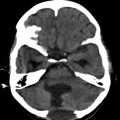
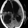
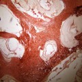
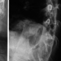
The disease usually spreads to the spine by direct extension from pulmonary, abdominal, or pelvic infestation and less often begins primarily in the vertebral body, affecting predominantly the dorsal region of the spine (Bhojraj and Shetty 1999; Garcia-Vicuna et al. 2000; Govender et al. 2000; Hamdan and Al-Kaisy 2000; Schnepper and Johnson 2004; Herrera et al. 2005; Prabhakar et al. 2005; Sapkas et al. 2006; Papanikolaou 2008; Spies et al. 2008). When hydatidosis begins in the spine, it usually begins in the marrow of a vertebral body and may progress around the neural arch, through adjacent ribs, reaching the spinal canal or the intervertebral discs; thus, spinal involvement is believed to occur through direct portovertebral venous shunts (Morshed 1977


Related posts:
Stay updated, free articles. Join our Telegram channel
Full access? Get Clinical Tree
