Hydronephrosis and obstructive uropathy


1. Hydronephrosis and obstructive uropathy


Introduction


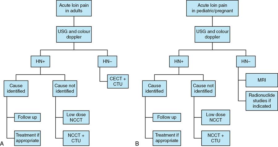
Hydronephrosis refers to the dilatation of renal pelvis and calyces. It can result from both obstructive and nonobstructive pathologies in the urinary tract. Obstructive hydronephrosis is more commonly encountered and urinary tract calculi represent the most common cause of obstruction in adults. Hydronephrosis is commonly associated with some amount of ureteric dilatation (hydroureter); however, isolated hydronephrosis may be seen when the obstruction is at the level of pelviureteric junction. Obstructive uropathy refers to the structural and functional abnormalities in the urinary system resulting from obstruction. It has been observed that obstructive nephropathy can rarely present without hydronephrosis. Patients with obstruction are prone to develop urinary infections and renal failure. Hence early diagnosis is essential to prevent irreparable damage to the urinary tract.


Aetiology


Obstructive hydronephrosis: The causes of obstructive dilatation can be categorized based on the level of obstruction and are enumerated in Box 10.12.3.1.1.


  1. A) Obstruction at the level of the ureter


    • Congenital – Stricture, ureterocele, pelviureteric junction obstruction
    • Acquired


      1. 1. Intraluminal


        • Calculus, blood clot, sloughed renal papilla, fungal ball, foreign material

      2. 2. Intramural


        • Neoplasms including epithelial and nonepithelial tumours, metastasis
        • Infective/Inflammatory lesions like TB ureteritis, schistosomiasis, ureteritis cystica, malacoplakia, leucoplakia, radiation ureteritis

      3. 3. Extrinsic


        • Hydronephrosis of pregnancy
        • Retroperitoneal tumours like lymphoma, sarcoma
        • Pelvic tumours like carcinoma cervix, ovary, rectum
        • Gynaecological causes like endometriosis, pelvic inflammatory disease, uterine prolapse, hydrocolpos
        • Gastrointestinal conditions like Crohn’s disease, diverticulitis, appendicitis, pancreatic and GI malignancy
        • Miscellaneous causes include retroperitoneal fibrosis, pelvic lipomatosis

  2. B) Obstruction at the level of urinary bladder and urethra


    • Congenital – Posterior urethral valve, hypospadias
    • Acquired


      1. 1. Benign conditions like benign prostatic hypertrophy (BPH), neurogenic bladder
      2. 2. Neoplasms include carcinoma of bladder and prostate


BOX 10.12.3.1.1


CAUSES OF OBSTRUCTIVE HYDRONEPHROSIS


OBSTRUCTION AT THE LEVEL OF URETER
Congenital Acquired
Stricture Intraluminal
Ureterocele Calculus
Pelviureteric junction obstruction Blood clot

Sloughed renal papilla

Fungal ball

Foreign material

Intramural

Neoplastic

Epithelial and nonepithelial tumours

Metastasis

Infective/Inflammatory

TB ureteritis

Schistosomiasis

Ureteritis cystica

Malacoplakia

Leucoplakia

Radiation ureteritis

Extrinsic

Hydronephrosis of pregnancy

Retroperitoneal fibrosis and tumours

Pelvic lipomatosis

Pelvic tumours

Gynaecological causes

Gastrointestinal conditions
OBSTRUCTION AT THE LEVEL OF URINARY BLADDER AND URETHRA
Congenital Acquired
Posterior urethral valve Benign
Hypospadias Benign prostatic hypertrophy

Neurogenic bladder

Neoplastic

Carcinoma bladder

Carcinoma prostate

Nonobstructive hydronephrosis (enumerated in Box 10.12.3.1.2):


  1. 1. Postobstructive dilatation
  2. 2. Vesicoureteric reflux
  3. 3. Diabetes insipidus
  4. 4. Congenital megacalyces
  5. 5. Primary megaureter
  6. 6. Polycalycosis


BOX 10.12.3.1.2


CAUSES OF NONOBSTRUCTIVE HYDRONEPHROSIS


  • Postobstructive dilatation
  • Vesicoureteric reflux
  • Diabetes insipidus
  • Congenital megacalyces
  • Primary megaureter
  • Polycalycosis

Pathophysiology


The changes in the renal structure and function depend on the level, extent and duration of the obstruction within the urinary tract.


  • Acute obstruction:


    • There is an increase in pressure within the collecting system proximal to the level of obstruction. Initially, there is dilatation of afferent arterioles due to the release of prostaglandins resulting in a transient increase in renal blood flow. However, 6–12 hours later, there is an increase in vascular resistance causing afferent arteriolar constriction and a decrease in renal blood flow. The rise in renal vascular resistance leads to a reduction in diastolic flow ending in organ ischaemia. There is atrophy of renal tubules secondary to raised intrarenal pressure resulting in reduced glomerular filtration rate (GFR). If the obstruction is relieved early, the renal function returns to normal subsequently. Irreversible renal damage sets in after 5–7 days and can progress to complete loss of function over a period of 6–8 weeks.

  • Chronic obstruction:


    • Chronic obstruction in the urinary tract leads to both structural and functional changes in the renal parenchyma. Long-term increase in intrarenal pressure causes diffuse cortical loss and renal parenchymal thinning. The kidney contracts in size with the deterioration of renal function. Partial obstruction may not result in significant functional impairment for years in few patients.

  • Hydronephrosis of pregnancy:


    • The majority of pregnant women develop moderate hydronephrosis and hydroureter involving the proximal two-thirds of the ureter in late pregnancy. The changes are usually more prominent on the right side due to compression of the right ureter by the enlarging uterus at the level of the pelvic brim. It is also attributed to an increase in maternal progesterone during pregnancy resulting in relaxation of smooth muscles of the ureters. It generally normalizes a few months postpartum.

Clinical features


Urinary obstruction may present with nonspecific symptoms. Loin pain is usually the most common presentation in acute urinary obstruction. Renal and ureteric pathologies result in referred pain in the loin region. It may be associated with renal angle tenderness, nausea, vomiting and microscopic haematuria in calculus disease. Sometimes renal failure may be the initial presentation in patients with underlying disease or sepsis. Patients with chronic obstruction may remain asymptomatic.


Diagnostic imaging


Imaging in patients with acute loin pain is aimed at not just identifying the cause of obstruction, but also the level and extent of obstruction and potential complications. The role of intravenous urography (IVU) as the primary imaging modality in suspected urinary obstruction has been superseded by the development of low dose noncontrast CT (NCCT) examination. The main advantage of IVU was the ability to determine both anatomic and functional alterations in the kidney secondary to obstruction. However, IVU requires serial delayed images to assess renal function which is cumbersome in an acute setting. Contrast-induced nephropathy is another potential concern with IVU which can be overcome using renal ultrasound and NCCT. CT urography has emerged as the imaging technique of choice in the evaluation of acute urinary obstruction due to both structural and functional assessment. The algorithm for the evaluation of patients with acute loin pain is depicted in the flow chart (Fig. 10.12.3.1.1).


Image
Fig. 10.12.3.1.1 An algorithm for the evaluation of patients with acute loin pain – (A) adults and (B) paediatric/pregnant patients.

Plain radiograph


A supine radiograph of the kidney, ureter and bladder (KUB) region can identify radiopaque calculi. Most of the calculi are calcium-containing and radiopaque. Radiolucent calculi containing uric acid, xanthine and other metabolic constituents are missed.


Intravenous urography


Acute obstruction can be diagnosed on IVU from the following signs (Fig. 10.12.3.1.2):


  1. 1. Dense persistent nephrogram – Nephrogram can last for 24 hours with a peak density at 6–8 hours when the obstruction is severe and the renal function is maintained. Significant functional impairment results in a lack of opacification of the collecting system.
  2. 2. Delayed pyelogram – It is essential to obtain delayed images until the level of obstruction is determined or it is concluded that contrast excretion is inadequate for opacification. The ‘rule of six’ for taking delayed images states that in the absence of contrast excretion on the 15-minute film, the next film must be obtained at least 6× the time interval between contrast injection and the last film. This minimizes unnecessary radiation exposure to the patient. Prone or dependent oblique images may be acquired to facilitate contrast excretion in patients with obstruction.
  3. 3. Enlarged kidney – Increase in size of the obstructed kidney as compared to the normal side.
  4. 4. Dilatation of pelvicalyceal system and ureter – This depends on the degree of obstruction. Blunting of the fornices is seen in mild cases, whereas loss of papillary impressions and ballooning of the calyces is seen with more severe cases.
  5. 5. Spontaneous pyelosinus extravasation – High-grade obstruction can result in rupture of fornices and extravasation of contrast. Smudging of calyces is seen with mild contrast leak. Profound extravasation is seen as contrast coursing along the renal pelvis and the psoas muscles to delineate the ureter with resultant urinoma formation.

Only gold members can continue reading. Log In or Register to continue

Stay updated, free articles. Join our Telegram channel

Mar 15, 2026 | Posted by in OBSTETRICS & GYNAECOLOGY IMAGING | Comments Off on Hydronephrosis and obstructive uropathy

Full access? Get Clinical Tree

Get Clinical Tree app for offline access