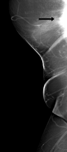
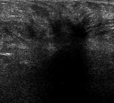
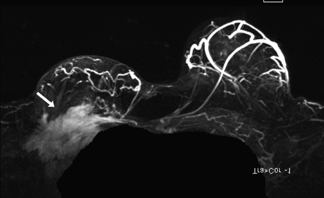
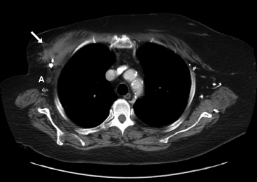
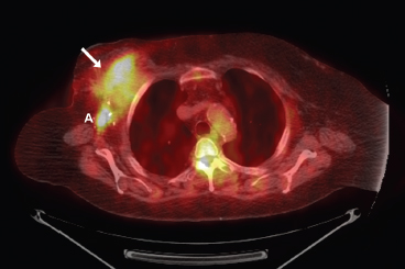
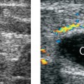
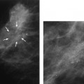
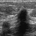
37 Identifications of Recurrent Disease A 65-year-old woman presents for MRI with a new palpable right chest wall mass and shoulder pain for 4 months. She has had a right modified mastectomy for breast cancer 17 years prior to this examination. Her axillary dissection revealed 3 of 17 nodes with metastatic tumor. She is also status post–right total shoulder arthroplasty 4 years prior to exam. MRI demonstrates a chest wall mass, so mammograms, right breast sonogram, and positron emission tomography–computed tomography (PET-CT) are performed. • Patient has a hard right chest wall mass. Fig. 37.1 Right axillary mammogram. There is an ill-defined, irregular mass (arrow) in the axilla. Frequency (Fig. 37.2) • 14 MHz Fig. 37.2 Right chest sonogram. The palpable chest wall mass corresponds to a heavily shadowing, spiculated, hypoechoic solid mass. Fig. 37.3 Contrast-enhanced bilateral breast MRI maximum projection intensity (MIP) image. There is an irregular right chest wall mass (arrow) that exhibits rapid enhancement and washout. Fig. 37.4 Axial positron emission tomography–computed tomography (PET-CT). There is 18F-fluorodeoxyglucose (FDG) uptake in the chest wall mass (arrow) that invades the pectoralis muscle. This exam also shows abnormal uptake in the right axillary nodes (A). Fig. 37.5
Case 37.1: Identification of Recurrent Disease
Case History
Physical Examination
Mammogram (Fig. 37.1)
Ultrasound
Other Modalities: MRI and PET-CT (Figs. 37.3, 37.4, and 37.5)
![]()
Stay updated, free articles. Join our Telegram channel


Full access? Get Clinical Tree


