
Image-Guided Brain Biopsy
Over the past two decades, image-guided needle biopsy has become an indispensable tool in modern neurosurgical practice.1–8 Just as image-guided stereotactic brain biopsy using reference frames largely replaced freehand techniques, biopsy using contemporary surgical navigation systems is gradually displacing frame technologies. This chapter reviews the principles of stereotactic needle biopsy, as well as advanced methodologies.
 Image-Guided Freehand Stereotactic Brain Biopsy
The advent of computed tomography (CT) in the 1970s marked the beginning of the modern era of brain biopsy. Surgeons could for the first time directly “see” the lesion they wished to sample on an image rather than infer its location by displacement of cerebral vessels or ventricular structures. Early efforts to exploit this information generally relied on “freehand” methods, often performed in the CT suite.9–11 A burr hole would be strategically placed and a biopsy instrument, sometimes just an angiocatheter, would be advanced to the target area while intermittently obtaining scans to check on its proximity to the target. On the rare occasion when the trajectory was within the plane of the image plane, this could be a relatively simple procedure, although the approach may not have been optimal with regard to intervening structures. When the approach crossed image slices, however, this could be a daunting, time consuming, and often hazardous procedure. Although the technique still has its advocates,9,12 most practitioners were eager to adopt a technique that offered unambiguous, rigid guidance to the lesion.
 Image-Guided Frame Stereotactic Brain Biopsy
In the late 1970s, it became recognized that CT scans harbored spatial information that could be mathematically extracted and applied to existing (or newly developed) stereotactic frames.5,13 This process usually required that a frame would be rigidly applied to the patient’s head prior to imaging, and would remain in place throughout the entire procedure, literally serving as a “frame of reference” for all that followed. Some type of adapter that served as a CT scan encoder was then secured to the frame in a stereotypical manner (Fig. 13–1), producing reference marks, or “fiducials,” on each image slice. The pattern of fiducials on the slice of interest could then be decoded to derive a coordinate, usually in Cartesian space (i.e., x, y, z) with respect to the frame. A guidance jig could then be set to direct the biopsy instrument (or other device) to that coordinate, when affixed to the reference frame (Fig. 13–2). Image-guided frame stereotactic brain biopsy (IGFSBB) set new standards for low morbidity and diagnostic success for brain biopsy.1,4,14–15 Also, the rationales for targeting and trajectory selection were established, better biopsy instruments developed, means of managing hemorrhage devised,16 and target imaging extended to magnetic resonance imaging (MRI) and other imaging techniques.17
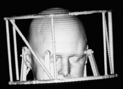
FIGURE 13–1. Three-dimensional surface rendering of Brown-Roberts-Wells (BRW) stereotactic localizer (Radionics, Burlington, MA), used to encode CT scans for stereotactic localization.
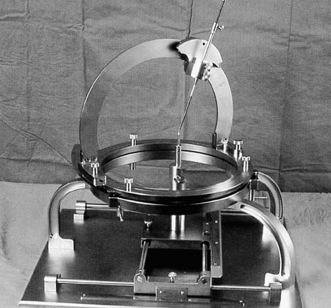
FIGURE 13–2. Brown-Roberts-Wells (BRW) frame atop its phantom.
Despite the clear advance over freehand techniques,11 IGFSBB was practiced by only a small fraction of neurosurgeons and became a niche procedure. Several reasons probably accounted for this, including that stereotactic frames were foreign to most practicing neurosurgeons and that the procedures were often logistically cumbersome, requiring frame application and imaging prior to the biopsy later that day, or use of invasive frames capable of reapplication.
 Image-Guided Frameless Stereotactic Brain Biopsy
Almost a decade after the development of IGFSBB, three technological developments occurred that, together, allowed for accurate neurosurgical guidance without reliance on a stereotactic reference frame: (1) the three-dimensional (3-D) spatial accuracy of both CT and MRI substantially improved, allowing for volumes, not just slices, of image data to be used for guidance; (2) inexpensive, spatially accurate 3-D digitizers were becoming available using sonic or multiarticulated arm technologies; (3) high-speed, reasonably inexpensive computers became available that could accurately present the location and orientation of a pointing device on displays of the image volume data.18–20
Related posts:

Stay updated, free articles. Join our Telegram channel

Full access? Get Clinical Tree


