, Maria Custódia Machado Ribeiro2 and Bruno Beber Machado3
(1)
Hospital da Criança de Brasília – Jose Alencar, Clínica Vila Rica, Brazília, Brazil
(2)
Hospital da Criança de Brasília – Jose Alencar, Hospital de Base do Distrito Federal, Brasília, Brazil
(3)
Clínica Radiológica Med Imagem, Unimed Sul Capixaba, Santa Casa de Misericordia de, Cachoeiro de Itapemirim, Brazil
Abstract
The spine of children and adolescents is frequently affected by diseases, some of them inherent to skeletally immature patients, such as juvenile idiopathic arthritis and Scheuermann’s disease, and others that are nonspecific of the pediatric group but present peculiarities when children are affected, such as isthmic spondylolisthesis and spondylodiscitis. Awareness of the imaging presentation of these conditions is paramount to the differential diagnosis and to guide therapeutic decision making. Some musculoskeletal disorders that present spinal involvement are discussed elsewhere in this book and will not be discussed in this chapter.
11.1 Introduction
The spine of children and adolescents is frequently affected by diseases, some of them inherent to skeletally immature patients, such as juvenile idiopathic arthritis and Scheuermann’s disease, and others that are nonspecific of the pediatric group but present peculiarities when children are affected, such as isthmic spondylolisthesis and spondylodiscitis. Awareness of the imaging presentation of these conditions is paramount to the differential diagnosis and to guide therapeutic decision making. Some musculoskeletal disorders that present spinal involvement are discussed elsewhere in this book and will not be discussed in this chapter.
11.2 Juvenile Idiopathic Arthritis
The spine is involved in a significant percentage of the patients with juvenile idiopathic arthritis (JIA), mostly in the upper cervical segment (facet joints) and in the craniovertebral junction. Similar to peripheral JIA, vertebral disease is related to synovial inflammation/hyperemia, pannus formation, and osteocartilaginous destruction. Radiographs are limited in the assessment of the arthritic spine due to the complex anatomy of this region, superimposition of bone structures, and low sensitivity to soft-tissue abnormalities. Computed tomography (CT) and magnetic resonance imaging (MRI) are the most useful imaging methods in spinal JIA: MRI is very suitable to evaluate the spinal cord and the nerve roots and to estimate the activity of inflammation, while CT is quite appropriate for bone and joint assessment.
Arthritic facet joints show narrowing of the joint spaces and erosions of the articular surfaces, with post-contrast enhancement in the inflamed tissues (Figs. 11.1 and 11.2); when present, ankylosis is found mainly in C2–C3 (Figs. 11.2, 11.3, 11.4, and 11.5). Synovitis of the atlantoaxial joint may lead to erosions of the odontoid peg, joint laxity, and vertebral instability. On radiographs, an interval of more than 5 mm between the posterior cortex of the anterior arch of C1 and the anterior cortex of the odontoid is considered abnormal (Figs. 11.1 and 11.6); lateral views of the cervical spine in flexion and extension are useful for assessment of instability. Additional findings include sub-axial subluxation (between the second and the seventh cervical vertebrae, also related to arthritis of the facet joints) and basilar invagination (related to occipitoatlantoaxial arthritis). Calcification of the soft tissues and of the vertebral ligaments is common, as well as reduced height of disk spaces (Figs. 11.3 and 11.4). Squaring and hypoplasia of the cervical vertebrae, related to chronic inflammation, are typical of long-standing JIA (Fig. 11.4).
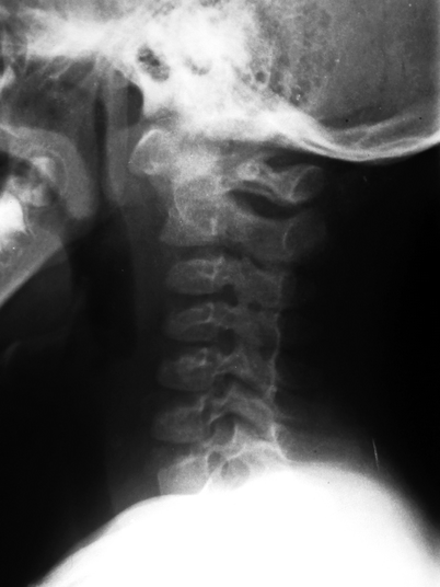
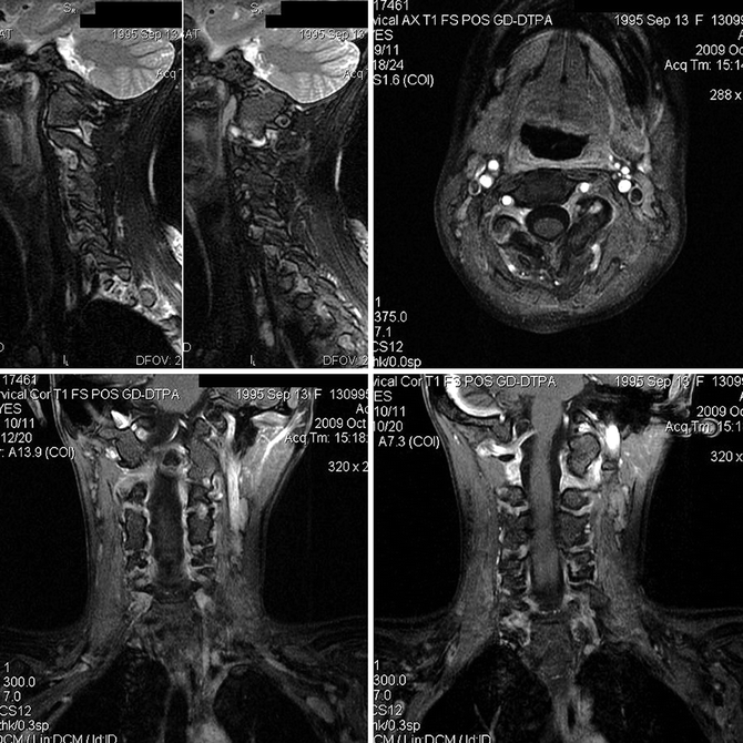
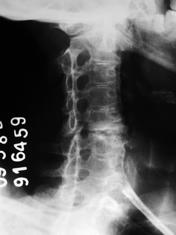
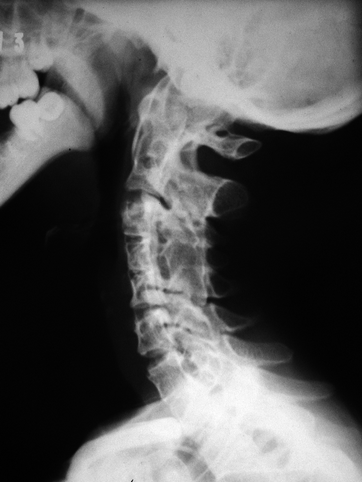
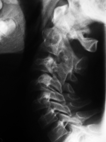
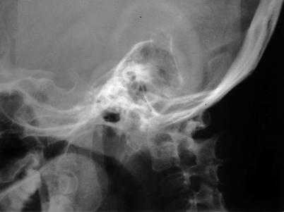
Fig. 11.1
Young child with active JIA in the cervical spine. Lateral radiograph disclose irregularity of the facets from C2 to C5, related to erosive joint disease, with minimal anterior slippage of C2. There is also atlantoaxial instability, with an increased interval between the anterior arch of C1 and the odontoid peg
Fig. 11.2
Contrast-enhanced MRI of the cervical spine of a 13-year-old female with active JIA. Sagittal fat sat T2-WI (upper-left image) and post-gadolinium fat sat T1-WI in the transverse (upper-right image) and coronal (lower row) planes reveal ankylosis of the facet joints in C4–C5. Erosions and post-contrast enhancement are seen in C2–C3 and C3–C4 at left and in C5–C6, bilaterally (Courtesy of Dr. Christiane Maria França Coimbra, Hospital Universitario de Brasilia – UnB, Brasilia, Brazil)
Fig. 11.3
Oblique radiograph of the cervical spine of a patient with long-standing JIA reveals extensive ankylosis of the facet joints. However, there is sparing of C5–C6 level, in which premature and atypical spinal degeneration can be seen, related to excessive solicitation of the only cervical segment with preserved mobility. Calcification of the nucleus pulposus is present in several disks, notably from C2 to C5
Fig. 11.4
Classic radiographic findings of long-standing spinal JIA. There is vertebral hypoplasia from C3 to C7, with squaring of the vertebral bodies and facet joint ankylosis, the latter more evident between C3 and C5. Ankylosis of the median atlantoaxial joint is also present, as well as reduced height of the disk spaces and peripheral disk calcifications from C3–C4 to C5–C6
Fig. 11.5
Ankylosis of the facet joints in C2–C3 can be seen in this lateral view of the cervical spine of a child with JIA. Note the normal appearance of the facet joints below this level, with regular and smooth surfaces, in contradistinction to those seen in the child of Fig. 11.1
Fig. 11.6
Lateral view (hyperflexion) of the craniovertebral transition of a patient with JIA demonstrating C1–C2 instability, with anterior translation of the atlas relative to the odontoid
11.3 Bacterial (Pyogenic) Spondylodiscitis
Discitis and vertebral osteomyelitis are different facets of the same process, namely, spondylodiscitis, which is more frequently found in the lumbar spine. Discitis, which is one of the ends of this spectrum, is more common in children from 6 months to 5 years old. Predominance of primary discitis in small children is probably due to trapping of septic emboli in the vessels of the cartilaginous portion of the disk, as these vessels tend to disappear with growth. On the other hand, vertebral osteomyelitis is the spinal counterpart of classic metaphyseal osteomyelitis, corresponding to up to 2 % of all cases of bone infection. It occurs mainly in older children (around age 7), resulting from hematogenous spread or from contiguous dissemination. Pyogenic infection of the spine is an aggressive and rapidly destructive process that may lead to acute neurologic damage and long-term complications. Timely diagnosis – before radiographic changes appear – is critical in order to start antibiotic therapy as soon as possible.
Imaging is usually necessary to confirm the clinical hypothesis of vertebral infection. Pyogenic spondylitis typically involves two adjacent vertebral bodies and the disk in between; this pattern of involvement appears on radiographs as reduced height of the affected disk space, ill-defined vertebral endplates, and bone erosions, which evolve very quickly once they become evident (Figs. 11.7 and 11.8). Even though this constellation of findings is virtually diagnostic in a child with strong clinical suspicion, it may take from 2 to 6 weeks for them to become apparent, and this delay renders radiographs inadequate for the assessment of initial disease. Bone scintigraphy becomes positive early in the course of the spinal infection, but MRI has similar sensitivity and superior resolution, being the latter the method of choice for early diagnosis. There is bone marrow edema adjacent to the affected endplates, with bone erosions and loss of cortical definition (Figs. 11.9 and 11.10). The affected disk presents reduced height, heterogeneous signal intensity, and post-gadolinium enhancement, the latter also found in the edematous bone marrow (Figs. 11.9 and 11.10). Intravenous gadolinium is indispensable, as the inflamed tissues enhance on post-contrast images: this feature is very helpful to estimate the real extent of the soft-tissue component, mostly paravertebral and epidural phlegmons and/or abscesses (Figs. 11.9, 11.11, and 11.12). Abscesses appear as well-delimited fluid collections with low signal intensity on T1-WI, high signal intensity on T2-WI, and peripheral post-contrast enhancement, while phlegmons are ill-defined areas of altered signal intensity with diffuse post-gadolinium enhancement (Figs. 11.9, 11.11, and 11.12). In general, the walls of bacterial abscesses are thicker and more irregular if compared to tuberculous abscesses. MRI is also useful to monitor treatment response, given that regression of soft-tissue abnormalities and fatty replacement of the formerly edematous bone are reliable indicators of cure. Nowadays, contrast-enhanced CT is a second-line method in the evaluation of pyogenic spondylodiscitis in children, as its diagnostic accuracy is lower than that of contrast-enhanced MRI (Figs. 11.12 and 11.13), being used essentially as a diagnostic alternative when the latter is not available or contraindicated. Gas bubbles or gas-fluid levels virtually exclude the possibility of tuberculosis and are indicative of the bacterial nature of the infectious process.
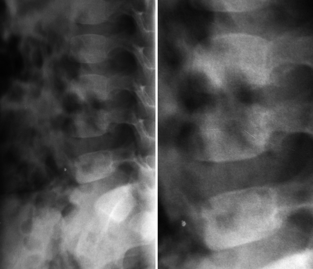
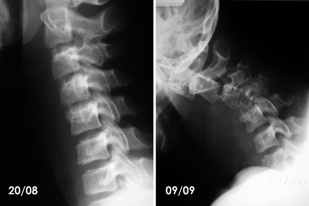
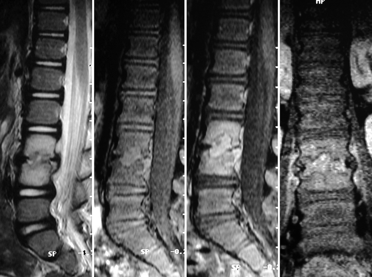
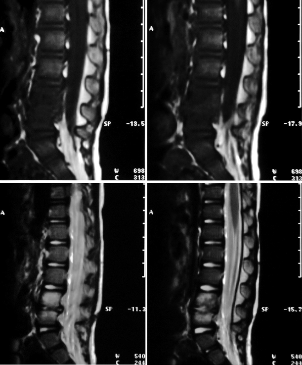
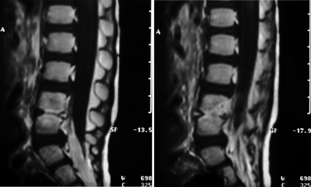
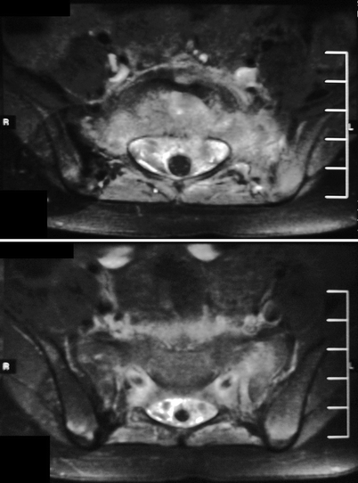
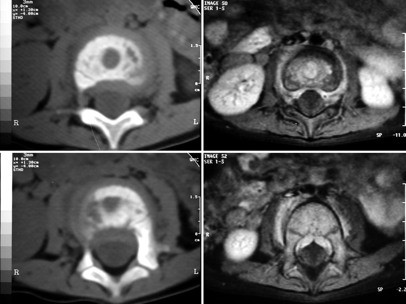
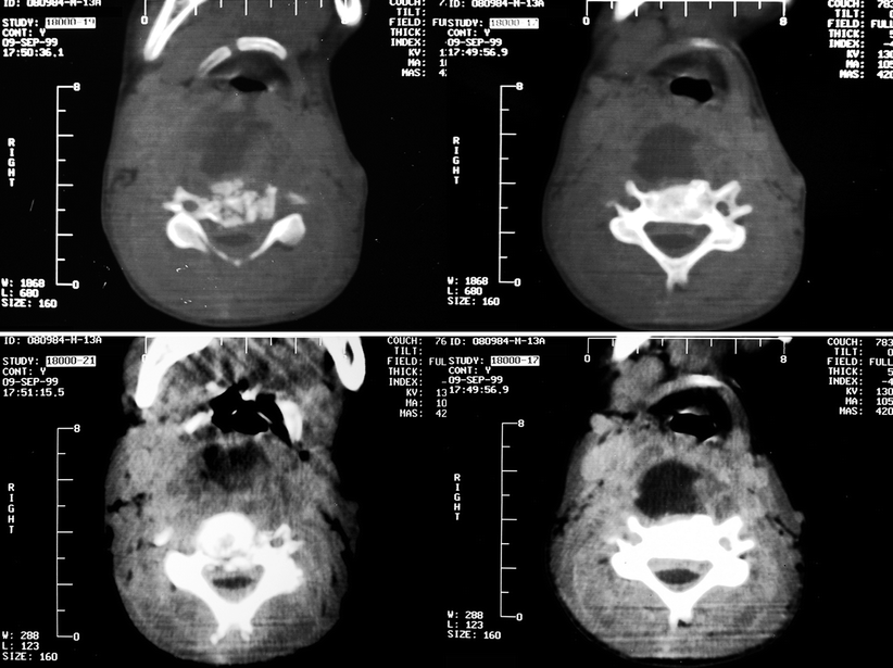
Fig. 11.7
L3–L4 pyogenic spondylodiscitis in a young child. Lateral radiograph reveals reduced height of the corresponding disk space, with loss of definition of the adjacent vertebral endplates (Courtesy of Dr. Marcelo Ricardo Canuto Natal, Hospital de Base do Distrito Federal and Pasteur Medicina Diagnostica – Diagnosticos da America S.A., Brasilia, Brazil)
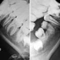
Fig. 11.8
Lateral radiographs of the cervical spine of a 13-year-old child with pyogenic spondylodiscitis, taken 20 days apart. The first radiograph (left) shows reduced height of C4–C5 disk space, with loss of definition of the upper endplate of C5 and swelling of the prevertebral soft tissues. The second radiograph (right) demonstrates rapid evolution of the infectious process, with angular deformity of the spine centered about C4 (related to bone destruction, collapse, and anterior wedging of this vertebral body), marked osteoporosis of the vertebral bodies from C3 to C5, and reduced height of the corresponding disk spaces. In addition, there is swelling of the prevertebral soft tissues leading to compression of the pharyngeal air column
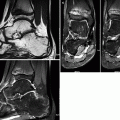
Fig. 11.9
Sagittal T2-WI (first image) and fat sat T1-WI (second image) of an 11-year-old female with pyogenic discitis in L3–L4 reveal reduced height and abnormal signal intensity of the affected disk, with destruction of the adjacent endplates and extension of the infectious process to the contiguous vertebral bodies. There is extensive bone marrow edema pattern surrounding the areas of bone destruction. A small subligamentous collection can be seen posterior to the vertebral body of L4. Sagittal (third image) and coronal (fourth image) post-gadolinium fat sat T1-WI demonstrate enhancement of the osteodiscal focus of infection, of the subligamentous abscess, of the areas of bone marrow edema, and of the edematous paravertebral soft tissues (Courtesy of Dr. Marcelo Ricardo Canuto Natal, Hospital de Base do Distrito Federal and Pasteur Medicina Diagnostica – Diagnosticos da America S.A., Brasilia, Brazil)
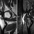
Fig. 11.10
L4–L5 pyogenic discitis. Sagittal T1-WI (upper row), T2-WI (central row), and post-contrast T1-WI (lower row) disclose diffuse reduction of the height of the affected disk, which presents abnormal signal intensity in all sequences. There is destruction of the adjacent endplates and bone marrow edema pattern. Post-gadolinium images demonstrate diffuse and intense enhancement of the infected disk and of the adjacent bone (Courtesy of Dr. Marcelo Ricardo Canuto Natal, Hospital de Base do Distrito Federal and Pasteur Medicina Diagnostica – Diagnosticos da America S.A., Brasilia, Brazil)
Fig. 11.11
Transverse post-gadolinium fat sat T1-WI of a 3-year-old child with L5–S1 bacterial spondylodiscitis. There is diffuse enhancement in the affected disk, in the bone marrow of the adjacent vertebrae, in the prevertebral and paravertebral soft tissues, and in the epidural space. Despite the extensive involvement of the epidural space, the absence of fluid collections and the diffuse – instead of circumscribed – appearance of such involvement favor phlegmon over abscess in this case
Fig. 11.12
Transverse CT images (left) and post-gadolinium fat sat T1-WI (right) acquired approximately at the same levels in a child with pyogenic spondylodiscitis of the upper lumbar spine. Despite the excellent resolution of CT for bone structures, allowing for accurate assessment of the destruction of the vertebral endplates, the evaluation of soft-tissue abnormalities is poor if compared to that obtained with MRI. In the latter, in addition to bone destruction and post-contrast enhancement of the bone marrow, there is also enhancement of epidural and paravertebral soft tissues, extending to the foramina, surrounding the emergent nerve roots and leading to anterior compression of the thecal sac (Courtesy of Dr. Marcelo Ricardo Canuto Natal, Hospital de Base do Distrito Federal and Pasteur Medicina Diagnostica – Diagnosticos da America S.A., Brasilia, Brazil)
Fig. 11.13
 Legg-Calvé-Perthes Disease
 Peculiar Aspects of the Anatomy and Development of the Growing Skeleton
 Sports-Related Musculoskeletal Lesions in Pediatric Patients
 Juvenile Idiopathic Arthritis
 Juvenile Spondyloarthropathies and Pediatric Collagen Vascular Disorders
 Accidental and Non-accidental Articular Injuries in the Skeletally Immature Patient


Contrast-enhanced CT of the neck of the same patient of Fig. 11.8, performed on the same day of the second radiograph. There is obvious destruction of the body of C4 (left images), with bone fragments extending to the spinal canal. A large retropharyngeal abscess is also seen, displacing the pharyngeal air column (right images). In addition, there is obliteration of the cervical fat planes (with diffuse post-contrast enhancement) and increased density of the epidural space, leading to the circumferential compression and deformation of the thecal sac
Related posts:
Legg-Calvé-Perthes Disease
Peculiar Aspects of the Anatomy and Development of the Growing Skeleton
Sports-Related Musculoskeletal Lesions in Pediatric Patients
Juvenile Idiopathic Arthritis
Juvenile Spondyloarthropathies and Pediatric Collagen Vascular Disorders
Accidental and Non-accidental Articular Injuries in the Skeletally Immature Patient
Stay updated, free articles. Join our Telegram channel
Full access? Get Clinical Tree
