Modern liposuction increasingly incorporates imaging technologies that improve anatomical visualization during planning and treatment. Radiologic evaluation provides additional information about fat distribution, tissue thickness, and structural anatomy. These insights support procedural precision during minimally invasive body contouring procedures.
The relationship between imaging and cosmetic surgery has expanded across multiple specialties. Liposuction radiology reflects this shift toward greater anatomical detail before and during intervention. Surgeons evaluating abdominal liposuction often rely on imaging to assess fat compartments, detect structural abnormalities, and refine surgical strategy.
Radiologic guidance does not replace surgical expertise. Instead, it provides objective anatomical data that informs treatment decisions and contributes to more predictable liposuction results.
Radiologic Assessment Before Surgery
Preoperative evaluation traditionally relied on physical examination and clinical judgment. Imaging now adds measurable anatomical information that supplements those methods.
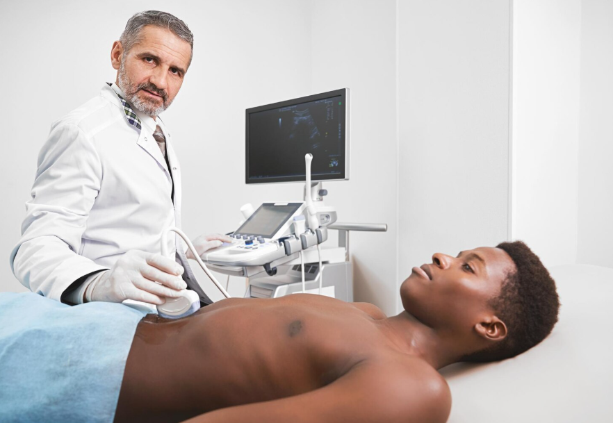
Ultrasound frequently serves as the primary imaging modality because it allows dynamic visualization of soft tissues without radiation exposure. CT and MRI may also be used in selected cases involving prior surgery or complex anatomy.
Radiologic imaging supports surgical preparation in several ways.
- Visualization of fat distribution across the abdomen, flanks, thighs, and submental region
- Identification of fibrotic tissue associated with previous procedures
- Detection of fluid collections such as seromas
- Recognition of structural abnormalities, including small abdominal wall hernias
- Measurement of adipose tissue thickness before treatment
These findings influence operative planning. Treatment zones can be mapped with greater accuracy. Cannula depth and trajectory are selected based on measurable tissue layers rather than surface assessment alone.
Radiologic evaluation also overlaps with other cosmetic specialties. Anatomical imaging plays a similar role in procedures involving facial cosmetic surgery, where tissue structure and postoperative changes are routinely analyzed through imaging.
Intraoperative Imaging And Procedural Precision
Imaging may also contribute during the procedure itself. Intraoperative ultrasound allows surgeons to observe adipose layers in real time. This capability improves depth awareness while suction is performed.
Consistent depth control influences final contour quality. Minor variations during suction can produce irregularities that become visible once swelling resolves.
Radiologic guidance during liposuction offers several practical advantages.
- Continuous monitoring of residual fat thickness
- Improved consistency between adjacent treatment zones
- Better navigation through areas affected by scar tissue
- Reduced likelihood of excessive fat removal in localized regions
These factors influence the stability of postoperative contour. Controlled removal produces smoother transitions across treated surfaces.
Patients researching cosmetic procedures often ask how safe liposuction is. Safety depends on multiple elements, including patient selection, anesthesia management, operative technique, and postoperative care. Imaging contributes by clarifying anatomy before and during treatment. Better anatomical awareness supports more controlled surgical execution.
Cost Structure And Imaging Considerations
Financial considerations remain an important part of cosmetic surgery planning. Patients frequently compare treatment options while researching the average liposuction cost.
Procedure pricing varies across clinical settings. Several variables influence the overall liposuction procedure cost.
- Number of treatment areas
- Procedure complexity
- Anesthesia requirements
- Facility and operating room fees
- Preoperative imaging and evaluation
- Postoperative monitoring
Imaging does not represent a universal requirement in every case. Its use depends on clinical judgment and anatomical considerations. Surgeons may recommend imaging when prior procedures, structural abnormalities, or uncertain tissue characteristics require additional evaluation.
As imaging technologies improve surgical planning and precision, patient education has also expanded. Individuals considering treatment frequently review how liposuction procedure costs are structured in modern clinics before selecting a personalized treatment plan.
Clinical Impact of Imaging on Liposuction Results
Radiologic imaging supports consistent body contouring outcomes through improved visualization of tissue layers. Mapping adipose compartments allows surgeons to perform more controlled fat removal while preserving smooth surface transitions.
The clinical roles of imaging during body contouring procedures can be summarized below.
| Imaging Function | Clinical Contribution | Influence On Outcomes |
| Preoperative fat mapping | Defines treatment zones and adipose depth | Promotes balanced contour removal |
| Scar and fibrosis detection | Identifies areas of increased resistance | Reduces contour irregularities |
| Fluid collection identification | Detects seromas and postsurgical changes | Improves treatment planning |
| Intraoperative ultrasound monitoring | Tracks remaining fat layer thickness | Prevents localized overresection |
| Postoperative imaging evaluation | Distinguishes swelling from complications | Clarifies recovery progression |
Radiologic interpretation also assists when postoperative symptoms require evaluation. Persistent swelling, asymmetry, or localized discomfort may prompt imaging to identify underlying causes.
This diagnostic role aligns with broader principles found in surgical radiography, where imaging supports both procedural planning and complication assessment.
Imaging in Postoperative Monitoring
Postoperative recovery following liposuction typically involves temporary swelling and bruising. Healing patterns vary depending on treatment area and procedural extent.
Imaging provides objective evaluation when recovery deviates from expected patterns. Ultrasound is frequently used because it allows rapid assessment of soft tissues.
Radiologic follow-up may help clinicians:
- Identify fluid collections that require aspiration
- Detect inflammatory changes or infection
- Evaluate tissue healing following large-volume liposuction
- Distinguish postoperative swelling from contour irregularities
Objective findings improve clinical decision-making. Imaging also supports communication between surgeons and patients during recovery. Visual evidence often clarifies the difference between normal healing and conditions requiring treatment.
Conclusion
Radiologic imaging has become an important component of modern liposuction procedures. Detailed visualization of tissue layers improves surgical planning and supports controlled fat removal. These capabilities contribute to procedural accuracy during minimally invasive body contouring.
Imaging also assists in postoperative assessment by clarifying recovery patterns and identifying complications when they occur. The integration of liposuction radiology therefore strengthens multiple stages of the treatment process, including planning, execution, and follow-up.
For further discussion of radiologic applications across medical and surgical practice, read more here
Related posts:
 Other Imaging Techniques in Dilated Cardiomyopathy
 Basic Echocardiography in Arrhythmogenic Right Ventricular Cardiomyopathy
 Early Diagnosis of Sports Injuries: Why Timely Imaging Matters
Enhancing Diagnostic Accuracy Through Online Radiology Consultation on Ai PACS
Dental Imaging Explained: From Bitewings To Panoramic X-Rays
Support Systems That Help Working Nurses Succeed in School
Early Diagnosis of Sports Injuries: Why Timely Imaging Matters
Enhancing Diagnostic Accuracy Through Online Radiology Consultation on Ai PACS
Dental Imaging Explained: From Bitewings To Panoramic X-Rays
Support Systems That Help Working Nurses Succeed in School
Stay updated, free articles. Join our Telegram channel

Full access? Get Clinical Tree


