, Maria Custódia Machado Ribeiro2 and Bruno Beber Machado3
(1)
Hospital da Criança de Brasília – Jose Alencar, Clínica Vila Rica, Brazília, Brazil
(2)
Hospital da Criança de Brasília – Jose Alencar, Hospital de Base do Distrito Federal, Brasília, Brazil
(3)
Clínica Radiológica Med Imagem, Unimed Sul Capixaba, Santa Casa de Misericordia de, Cachoeiro de Itapemirim, Brazil
Abstract
The last decades of the twentieth century and the beginning of the twenty-first century have witnessed unparalleled improvements in diagnostic imaging. Until the 1960s, plain radiographs were the only technique available in this field. In time, however, sophisticated imaging methods were gradually added to the diagnostic arsenal, and even radiographs have entered into the digital arena. Each of these imaging methods has singularities to be considered in the assessment of immature joints, and both the radiologist and the requesting physician must be familiar with them. The requesting physician is supposed to have an open-minded and receptive attitude, consulting the radiologist whenever a doubt arises (e.g., “which imaging method is the most appropriate for this patient in this specific clinical setting?”) and providing all the relevant information for each case. Accordingly, the radiologist must perform the requested imaging study and interpret the findings under the light of the clinical picture. Analyzing the images without taking the clinical background into account is dangerous and may lead to an erroneous – and potentially catastrophic – diagnosis.
1.1 Introduction
The last decades of the twentieth century and the beginning of the twenty-first century have witnessed unparalleled improvements in diagnostic imaging. Until the 1960s, plain radiographs were the only technique available in this field. In time, however, sophisticated imaging methods were gradually added to the diagnostic arsenal, and even radiographs have entered into the digital arena. Each of these imaging methods has singularities to be considered in the assessment of immature joints, and both the radiologist and the requesting physician must be familiar with them. The requesting physician is supposed to have an open-minded and receptive attitude, consulting the radiologist whenever a doubt arises (e.g., “which imaging method is the most appropriate for this patient in this specific clinical setting?”) and providing all the relevant information for each case. Accordingly, the radiologist must perform the requested imaging study and interpret the findings under the light of the clinical picture. Analyzing the images without taking the clinical background into account is dangerous and may lead to an erroneous – and potentially catastrophic – diagnosis.
This chapter is a brief introduction to the most important imaging methods used in the investigation of the immature joint, emphasizing their peculiarities in pediatric patients. It is important to keep in mind that these imaging methods are complementary instead of exclusive, acting in a synergistic way when properly combined.
1.2 Radiographs
Radiographs were always closely associated with the assessment of the locomotor apparatus and its disorders. It is emblematic that the first radiographic study ever made was the image of a hand, the left hand of the wife of Wilhelm Röntgen, the discoverer of the X-rays, in 1895. In its classic form, currently named “conventional” radiography, a radiographic film is placed inside a cassette with intensifying screens and exposed to X-rays. The anatomic structure under study is interposed between the source of the radiation and this cassette. The resulting image translates the different densities of the several body tissues: denser tissues, such as the bone, appear whiter as they absorb the radiation in a higher degree, therefore limiting the amount that effectively reaches the film. Similarly, less dense tissues, such as fat, appear darker. Even though digital radiographs – either obtained with computed radiography (CR) or digital radiography (DR) – take advantage of the same physical principles, they do not rely on X-ray films to produce medical images. On the contrary, a sensor receives the emitted radiation and transmits the electronically obtained information. The generated image is, in fact, a computer file, which can be viewed on the screen of a workstation, archived in many types of digital media, or printed (either on paper or on a medical imaging film). Despite the advantages of the digital modalities over the conventional technique (wider dynamic range, less exposure to radiation, telemedicine capabilities – Fig. 1.1), the training and the experience of the observer are far more important than the technology involved.
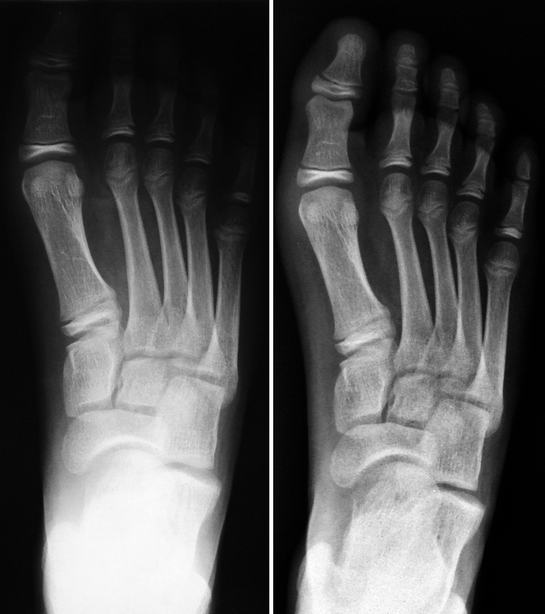
Fig. 1.1
Radiographs of the left foot performed with conventional (left) and digital (right) techniques. The digital radiograph has wider dynamic range if compared to the conventional one: the toes and the tarsal bones are equally well seen on the former, while only the midfoot is adequately exposed on the latter
The notion that X-rays became outdated after the advent of modern imaging methods has no grounds. Radiographs remain essential and consist in the first line of investigation for patients with articular disorders, so that they must be requested before soliciting any additional imaging study (Fig. 1.2). They are inexpensive and widely available, are not operator dependent, and present good reproducibility. Correct interpretation of imaging studies like ultrasonography and magnetic resonance imaging relies largely on correlation with radiographs. Two or more views are usually obtained, making it easier to determine the location of abnormal findings. Serial radiographs may be used to monitor the course of articular disease and are employed as imaging criteria in several staging and scoring systems (Fig. 1.2). In arthritic patients, for example, radiographs are very suitable for demonstration of established osteoarticular damage, such as cortical erosions, abnormal joint alignment, joint space narrowing, and ankylosis (Fig. 1.3). Moreover, radiographs are often problem-solving by themselves, as several musculoskeletal disorders can be readily diagnosed on X-rays (e.g., bone tumors, fractures, osteomyelitis, congenital malformations, and developmental abnormalities – Figs. 1.4 and 1.5). Disadvantages include the use of ionizing radiation (which is especially deleterious for the immature organism), low sensitivity for the early stages of articular diseases, and limited usefulness in the assessment of the soft tissues and anatomically complex joints. Conventional arthrograms and linear tomography (planigraphy) are radiographic techniques that were popular in the past but became obsolete after the advent of cross-sectional imaging, given the higher diagnostic accuracy and the noninvasive nature of the latter (Fig. 1.6).
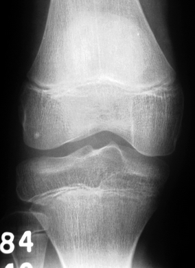
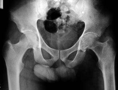
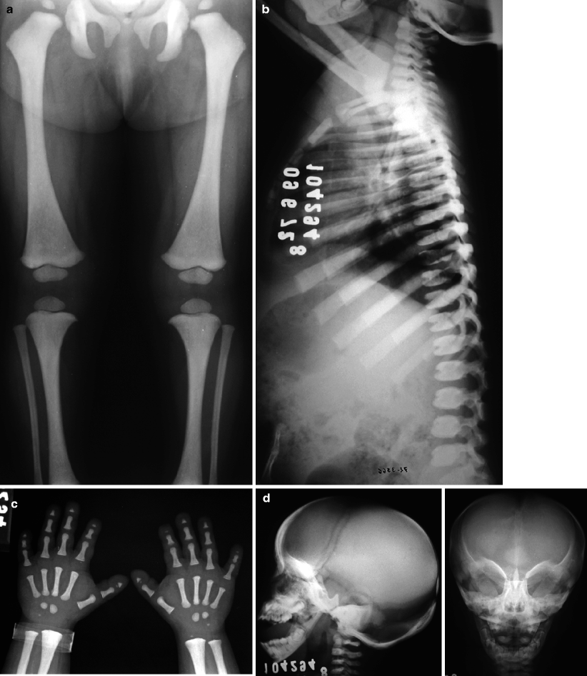
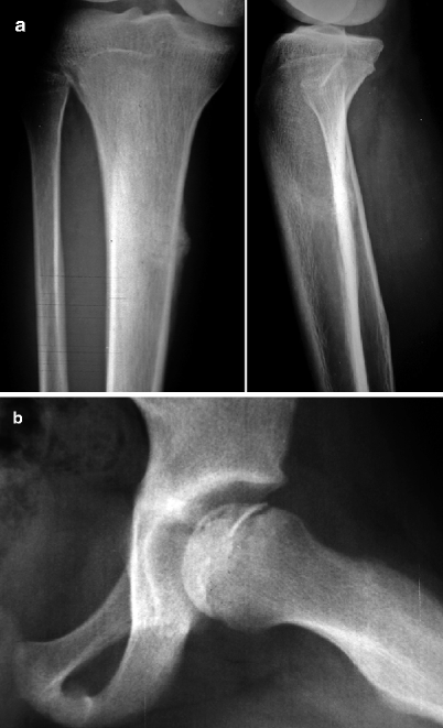
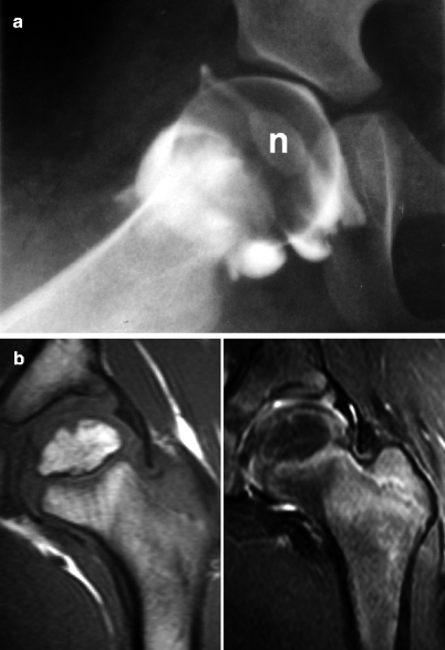
Fig. 1.2
Radiograph of the right knee of a 13-year-old female with juvenile idiopathic arthritis diagnosed one year before. Even though erosions are absent, osteoporosis and subtle increase in the size of the epiphyses are already seen, related to the hyperemic state. This radiograph will serve as a baseline study to monitor disease evolution and treatment response
Fig. 1.3
Pelvic radiograph of a 20-year-old male with juvenile idiopathic arthritis since age 15. There is advanced arthritis of the left hip, with regional osteoporosis, erosions of the articular surfaces, narrowing of the joint space, and reduced size of the homolateral femoral head, which is markedly deformed
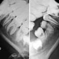
Fig. 1.4
Radiographs of a child with short stature showing generalized increase in bone density, with loss of definition of the corticomedullary junction (a, b). The fingers are short and stubby and the distal phalanges are hypoplastic (c). Craniofacial abnormalities include macrocrania, wide sutures and fontanelles, parieto-occipital bossing, and straightening of the mandibular angles (d). These findings are typical of pyknodysostosis, with no need for additional investigation
Fig. 1.5
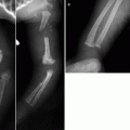
In an appropriate clinical background, some radiographic patterns are almost pathognomonic. However, in most cases these findings usually appear late in the course of the disease, limiting their effectiveness for early diagnosis. In a, there is a cortical lucency in the medial aspect of the proximal diaphysis of the right tibia associated with periosteal reaction and bone neoformation, which are highly suggestive of stress fracture in an adolescent runner. In b, radiograph of a child with pain in the left hip shows that the femoral head is sclerotic and reduced in size, presenting a curvilinear subchondral lucency (crescent sign), findings that are typical of Legg-Calvé-Perthes disease. In both cases, MRI would be able to establish the diagnosis much earlier
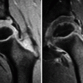
Fig. 1.6
Until the advent of modern imaging methods, only invasive radiographic procedures, such as conventional arthrograms (a), were able to demonstrate the epiphyseal cartilage, demanding sedation and intraarticular injection of iodinated contrast. This cartilage is much larger than the mineralized ossification nucleus (n), the only portion of the epiphysis that is detectable on radiographs. In (b), for comparison, coronal T1-WI (left) and fat sat T2-WI (right) of the left hip of a 3-year-old child are able to display both calcified and non-calcified structures with high detail and in a noninvasive way, including the epiphyseal cartilage, the acetabular labrum, muscles, and tendons
1.3 Ultrasonography
Ultrasonography (US) has become widely accepted for joint assessment, notably after the introduction of high-frequency transducers. In US, an array of piezoelectric crystals generates a beam of ultrasonic waves that is directed to a given structure, which will reflect them in different degrees, depending on the composition of its tissues. For example, the synovial fluid is a very good conductor of ultrasonic waves, while the cortical bone reflects them almost entirely. The reflections of the ultrasonic beam are transmitted to a computer that will process the obtained data and produce real-time images in a screen, therefore providing a unique dynamic characteristic to US. Although excellent for the evaluation of large joints and superficial soft tissues (Figs. 1.7 and 1.8), this study presents limitations in the assessment of deeply seated structures or small/deformed joints, to which the transducer may not fit satisfactorily. In addition, the ultrasonic beam relies on the existence of acoustic “windows” to reach the region of interest. Superficially located osseous abnormalities may occasionally be detected on US, but essentially no information is gathered about the innermost portions of the bones. Doppler US is a technique that detects and measures blood flow in vascular structures, being very useful in the investigation of hyperemic joint diseases and soft-tissue masses and fluid collections (Figs. 1.9 and 1.10).
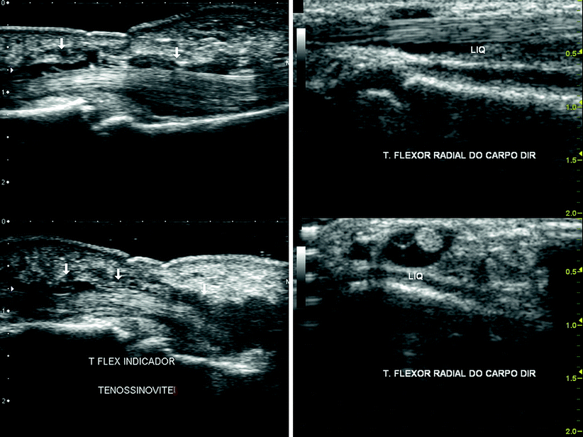
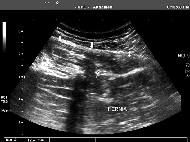
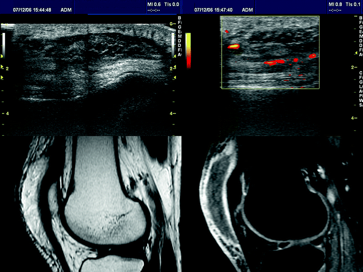
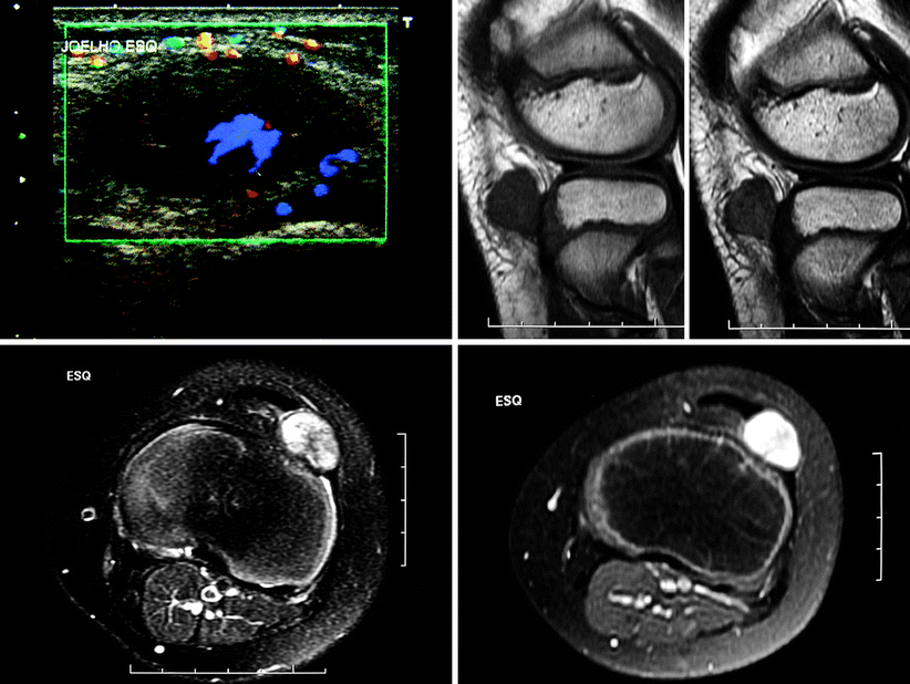
Fig. 1.7
US of two different patients with juvenile idiopathic arthritis and tenosynovitis involving the flexor tendons of the index finger in the first (left) and the flexor carpi radialis in the second (right). The tendons are elongated on sagittal scans (left images and upper-right image) and appear as rounded structures on transverse scans (lower-right image), with a fibrillar appearance. The synovial fluid in the tendon sheaths appears hypoechogenic, while amorphous echogenic foci are related to synovial thickening and debris. Even a 4-year-old child (first patient) tolerates an US scan very well
Fig. 1.8
US of an adolescent complaining about a nodule of elastic consistency and variable dimensions in the lumbar region. The image reveals a focal defect of the lumbar aponeurosis measuring 12 mm (calipers) and herniation of paravertebral muscle fibers
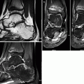
Fig. 1.9
An 18-year-old female with autoimmune hepatitis since age 14 and taking immunosuppressant drugs, presenting with left-sided pyogenic prepatellar bursitis. US scans (upper row) reveal thickening of subcutaneous tissue and filling of the prepatellar bursa with heterogeneous hypoechogenic material; peripheral hyperemia is seen on Doppler US (red-yellowish streaks in the second image). In the lower row, T2-WI (first image) discloses subcutaneous thickening and filling of prepatellar bursa with heterogeneous content, mostly hyperintense. Post-contrast fat sat T1-WI (second image) reveals enhancement of the synovium and of the inflamed subcutaneous tissue, corresponding to the hyperemic zones seen on Doppler. The non-enhancing material inside the bursa corresponds to debris and pus
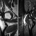
Fig. 1.10
A 9-year-old boy presenting with a painless infrapatellar lump in the anterolateral aspect of the left knee. Doppler US (upper-left image) discloses an oval hypoechogenic mass, well delimited, and heterogeneous, with central areas of increased blood flow (lavender). Sagittal T1-WI (upper-right image), transverse fat sat T2-WI (lower-left image), and post-gadolinium fat sat T1-WI (lower-right image) reveal a sharply delimited lesion of heterogeneous appearance, predominantly hypointense on T1-WI and hyperintense on T2-WI, whose epicenter is in the patellar tendon. There is marked post-gadolinium enhancement, as could be inferred from the increased flow seen on Doppler. The histopathological diagnosis was fibroma of the tendon sheath
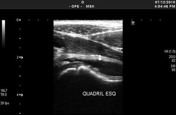
US is largely used in pediatric patients because it is painless, inexpensive, and widely available; can be performed at the bedside; and does not require special or uncomfortable positioning. The absence of ionizing radiation must also be taken into account in this age group, as serial studies can be performed without risk of damage to the growing organism. It is well tolerated by children, and several joints can be examined expeditiously in a single session. Joint effusion and synovial thickening are readily apparent on US, as well as extension of the inflammatory process to adjacent bursae, synovial sheaths, or cysts (Figs. 1.7, 1.9, and 1.11). The articular cartilage is particularly well seen in large joints (Fig. 1.11). In arthritic patients, the degree of hyperemia can be estimated with Doppler US and monitored in sequential studies, serving as an indicator of disease activity and a marker of treatment response. The dynamic properties of US allow for assessment of a given structure in different spatial planes (Fig. 1.7), evaluation of articular relationships (such as in newborns with suspected developmental dysplasia of the hip – Fig. 1.12), and real-time demonstration of abnormalities that would go unnoticed with other imaging methods, such as tendon dislocations or muscular hernias (Fig. 1.8). Additionally, US is very useful to guide invasive diagnostic and/or therapeutic procedures.
 Legg-Calvé-Perthes Disease
 Peculiar Aspects of the Anatomy and Development of the Growing Skeleton
 Sports-Related Musculoskeletal Lesions in Pediatric Patients
 Juvenile Idiopathic Arthritis
 Juvenile Spondyloarthropathies and Pediatric Collagen Vascular Disorders
 Accidental and Non-accidental Articular Injuries in the Skeletally Immature Patient


Related posts:
Legg-Calvé-Perthes Disease
Peculiar Aspects of the Anatomy and Development of the Growing Skeleton
Sports-Related Musculoskeletal Lesions in Pediatric Patients
Juvenile Idiopathic Arthritis
Juvenile Spondyloarthropathies and Pediatric Collagen Vascular Disorders
Accidental and Non-accidental Articular Injuries in the Skeletally Immature Patient
Stay updated, free articles. Join our Telegram channel
Full access? Get Clinical Tree
