Fig. 1
(a) 1: Common trunk; 2: left anterior descending artery; 3: intermediate branch. (b) Arrow: Circumflex artery
(a)
Left anterior descending artery ( LAD ): It courses anteriorly along the interventricular groove to the apex. From this artery originate:
The septal perforator arteries which supply the anterior two-thirds of the interventricular septum.
The diagonals, which supply the anterolateral portion of the left ventricle; they are up to six, and they are numbered sequentially as they arise (D1, D2 …).
Moreover, LAD can be divided into three main portions:
Proximal portion: from the origin to the first septal perforator or diagonal artery.
Mid portion: from the origin of the first septal perforator or diagonal artery to the second diagonal.
Distal portion: from the origin of the second diagonal up to the apex.
Occasionally, the LMA does not divide but trifurcates into LAD, LCX and a middle branch between them, known as ramus intermedius.
(b)
Left circumflex artery ( LCX ): It courses along the left atrioventricular groove up to the obtuse margin of the heart (in 80–85 % of patients, see below); it supplies the lateral portion of left ventricle. From this artery originates the obtuse marginal arteries (OM) numbered sequentially as they arise (OM1, OM2 …). It can be divided into two portions:
Proximal portion: from origin to the origin of the greatest OM
Distal portion: from the origin of the greatest OM.
There is another one important concept to explain: the dominance (Fig. 2). The dominance of the coronary circulation is determined by the origin of the atrioventricular nodal artery, PDA and PLV. In fact, the dominance can be [5]:
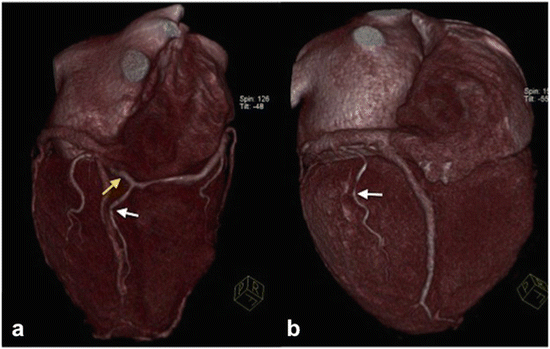

Fig. 2
(a) Right dominance: The posterolateral branch (yellow arrow) and the posterior interventricular branch (white arrow) originate from the right coronary artery. (b) Left dominance: The posterior interventricular branch (arrow) originates from the circumflex artery
1.
Right dominance (80–85 %): the atrioventricular nodal artery, PDA and PLV originate from RCA.
2.
Left dominance (8–10 %): the atrioventricular nodal artery, PDA and PLV originate from LCX.
3.
Codominance (7–8 %): the PDA originates from the RCA and the PLV originates from the LCX.
American Heart Association Left Ventricle Segmentation Model and Coronary Distribution Territories
According to the AHA left ventricle segmentation model [5, 6] we can divide the left ventricle cavity into three regions on the long axis, and the walls in 17 segments (seen on a short-axis plane and numbered in anticlockwise) as you can see in the table (Table 1) and in the figure (Fig. 3).
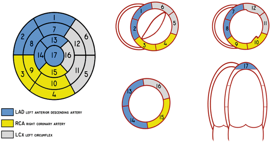
Table 1
AHA left ventricle segmentation model
Basal region | Mid-cavity | Apical region |
|---|---|---|
Anterior (1) | Anterior (7) | Anterior (13) |
Anteroseptal (2) | Anteroseptal (8) | Septal (14) |
Inferoseptal (3) | Inferoseptal (9) | Inferior (15) |
Inferior (4) | Inferior (10) | Lateral (16) |
Inferolateral (5) | Inferolateral (11) | Apex (17) |
Anterolateral (6) | Anterolateral (12) |

Fig. 3
AHA left ventricle segmentation model and blood supply
Regarding the blood supply, the LAD provides for the supply of blood of the segments 1, 2, 7, 8, 13 and 14; LCX provides for the supply of blood of the segments 5, 6, 11 and 12, meanwhile RCA (when it is dominant) supplies segments 3, 4, 9, 10 and 15 [6]. The segment 17 can be supplied by any of the three arteries, but generally is assigned to the LAD [6].
The Coronary Artery Calcium Score
The CACS is a method of evaluation of the amount of calcium localised in the coronary arteries, which is indicative of the risk of cardiac major events [7], but its use in clinical practice is controversial [8]. It has been introduced in the clinical practice with the invention of the electron beam scanners, and many authors have proposed different methods of measurement of this score.
The most important and tested in clinical practice is the so-called Agatston-Janowitz score [8, 9] introduced by Agatston and colleagues in 1990: it is a semi-quantitative method calculated on the basis of basal ECG-gated scans at 130 kV and 630 mA, with a slice thickness of 3 mm, on a matrix of 512 × 512 [8]. In order to be included in the calculation of the score, all the calcifications of the coronary arteries must cover a surface of more than 1 mm2 and have to have a value of at least 130 HU [8]. The area of every single calcification is then multiplied for an attenuation-weighting factor (with a value between 1 and 4) according to the highest HU value of the plaque [8]. In this way it is possible to calculate the calcium score of the single vessels, or the total calcium score given by the sum of the calcium vessels calcium score [8]. Nowadays various software which automatically calculate the CASC are available, in particular using the Agatston-Janowitz method.
The indications for the execution of the CT scan for the CASC are the evaluation of asymptomatic patients with an intermediate risk for CAD (values higher than 400 according to the Agatston-Janowitz score suggest modifications in the lifestyle and medical therapy in order to reduce the risk of major cardiac events), and of symptomatic patients as screening before invasive procedures or CTA (CTA could be useless in patients with high calcium score because of the increased incidence of artifacts), or if it is impossible to execute other examinations [8].
The CASC is directly related with the risk of major cardiac events, and it has been seen a gradual decrease of the overall survival related with the increase of the Agatston score, in particular for value above 1,000 [10].
Anyway, it is important to underline that the CASC does not allow the evaluation of the patency of the coronary arteries and the number, the morphology and the characteristics of the non-calcified plaques [8].
CTA Examination
Thanks to the introduction of single-source 64-slice CT scanner, CTA has become a very useful and feasible exam for the assessment of coronary arteries. In fact this kind of CT scanners, because of their high temporal (up to 150 ms) and spatial resolution, have higher sensitivity and specificities in CAD detection (sensitivity 85–99 % and specificity 86–96 %) despite 4-slice (sensitivity and specificity between 76 and 93 %) and 16-slice CT scanner (sensitivity and specificity between 82 and 95 %) [11]. In addiction, the introduction of 128-slice, 256-slice and 320-slice CT scanner in clinical practice and the dual source CT (DSCT) has further improved spatial and temporal resolution (up to 75 ms in DSCT), and allows to reduce radiation dose in combination with some dose-saving strategies (see below).
The great challenge of CTA is freezing a continuous moving structure, and the collaboration of the team radiologist-technician with the patient has to be as best as possible in order to make a diagnostic exam.
The anamnesis is a fundamental part of radiology (and of course in medicine), and in particular in CTA: The radiologist has to know every useful data of the patient for the correct interpretation of the examination. In particular the radiologist has to investigate on familiarity for CAD, previous and/or coexisting pathologies (diabetes, hypercholesterolemia, hypertension …), smoke, symptomatology (angina, dyspnea …) and possibly the results of previous tests executed (basal and stress electrocardiography, basal and stress echocardiography …); moreover, since CTA is an examination which involves the use of iodinate contrast medium, the radiologist must investigate about any risk factors concerning that (see Chap. “Principles of Computed Tomography”).
After the anamnesis, the radiologist has to explain to the patient the phases and the risks related to the procedure, obtaining the informed consent.
Once assured about this, another important step is the measurement of the heart beat rate (HBR): in patients with HBR >65 bpm the use of β-blockers could be necessary (in particular β1-selective blockers, such as metoprolol, atenolol) so as to lower HBR and to make the rhythm more regular [12]. We obtain better scans if the heart beat rate is <65 bpm, and this value is indispensable if a prospective ECG-triggering acquisition mode is chosen (for the use of β-blockers in transplanted heart see below). Contraindications to their use are seen in the table (Table 2) [12].
Table 2
Contraindications to β-blockers
Heart rate <60 bpm |
Systolic blood pressure <100 mmHg |
Uncompensated cardiac failure |
Aortic stenosis |
Allergy to β-blockers |
Asthma or COPD on β2-agonist inhaler |
Active bronchospasm |
Second- or third-degree atrioventricular block |
As these conditions are excluded, the radiologist could administer a premedication of β-blockers orally. For metoprolol, one 50 mg dose at least 1 h before the examination monitoring the HBR every 15 min: if it does not lower <65 bpm (or <60 bpm if the HBR is irregular) and after a 15-s breath hold there is not any lowering of the frequency, metoprolol I.V. can be administered when the patient lies on the moving table, one 2.5 mg dose in 1 min, and if necessary another 2.5 mg dose after 5 min; if the HBR still remains elevated after 5 min the last bolus could be given up to two additional single doses of 5 mg every 5 min (maximum I.V. dose: 15 mg) [12]. If metoprolol I.V. is given, the patient has to be observed at least 30 min after the last administration; if the HBR is <45 bpm, the radiologist has to consider the use of atropine, whereas if bronchospasm occurs it is necessary administering a β-agonist (for example albuterol) [12].
Patients with severe arrhythmias (such as atrial fibrillation) are very difficult to evaluate because of the irregular heart rhythm; further technologic improvements have still to be made [13].
Anxious patients have to be ensured, because anxiety is related with high HBR; if it would be necessary, it is moreover advised administering oral benzodiazepine such as diazepam 5 mg (5–15 drops 15 min before the examination).
After these preliminary moments, the patient can be placed supine on the moving table, with ECG electrodes positioned on his/her chest, and a cannula (minimum dimension 18G) inserted in the right antecubital vein. ECG is registered and analysed by the CT scanner in real time. Every CTA is achieved through ECG-gated scans .
The technician makes the scanogram, and then places a ROI on the aortic root: the scan delay time from the beginning of contrast medium I.V. infusion is chosen according to a bolus test (20 ml contrast medium bolus at the same flow of the examination) or an automatic bolus-triggering technique [5], knowing the technical specifics of the own CT scanner. The scan has to cover the chest from the diaphragm up to the aortic arch, in order to cover the heart and the great thoracic vessels. In certain cases, the scan has to cover also the epiarotic vessels (in particular in patients with arterial coronary artery bypass graft (CABG), see below). Few seconds before the beginning of the scan, it is recommended to administer nitroglycerine 0.4 mg sublingual (spray or tablet) to dilate the coronary arteries [14].
The next step is the contrast medium infusion: Generally a 50–60 ml contrast medium bolus is administered, with a biphasic (contrast medium bolus followed by a 50 ml saline solution bolus to wash the right chambers and better evaluate RCA) or triphasic technique (contrast medium bolus followed by a mixture of contrast medium and saline solution to evaluate right chamber, and followed then by a bolus of saline solution) [5], with a flow of 5–6 ml/s.
The acquisition mode depends on the type of scanner used; for a single-source CT scanner (64-slice CT scanner or more) two different techniques can be used (it is necessary that the patient holds the breath during the examination):
“Retrospective ECG gating” (Fig. 4): It is a spiral scan, with a very low pitch (0.2–0.4) [11], acquired continuously and simultaneously to the ECG [15]. Once scan is obtained, CT scanner elaborates the projection data acquired during the helical scan with the ECG, and reconstructs the exam “retrospectively”, obtaining images of the entire cardiac cycle. The images can be reconstructed in two different ways, with the partial scan reconstruction or the multiple-segment reconstruction (see below).
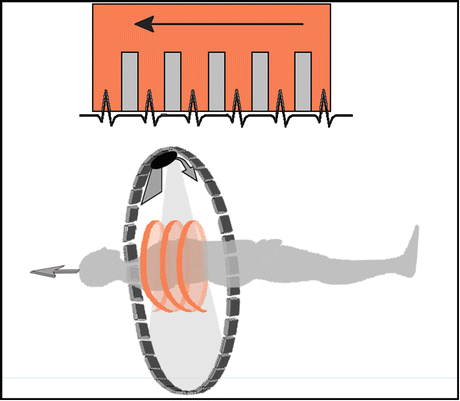
Fig. 4
Retrospective ECG gating: The scan is obtained with a spiral CT during the entire cardiac cycle and it is then retrospectively reconstructed according to the registered ECG
The advantages of this technique are a good temporal resolution (80–250 ms) [15] better than prospective ECG-triggering acquisition, whereas the absorbed dose is very high (between 7.6 and 31.8 mSv) [16] because of continuous radiation emission during the scan (very low pitch and great overlap in the same section of the thorax) [15].
“Prospective ECG triggering” (Fig. 5): Even in this case, the scan is made synchronised with the ECG. It is a sequential scan: data acquisition takes place only in a predefined temporal window of the R-R cycle (i.e., in the diastolic phase, when the heart is more stable, between 20 and 80 % of the R-R cycle; or between 50 and 80 % if the rhythm is lower, in order to reduce the amount of radiation dose), and the X-ray tube turns only when triggered by the ECG signal registered by the CT scanner [11].
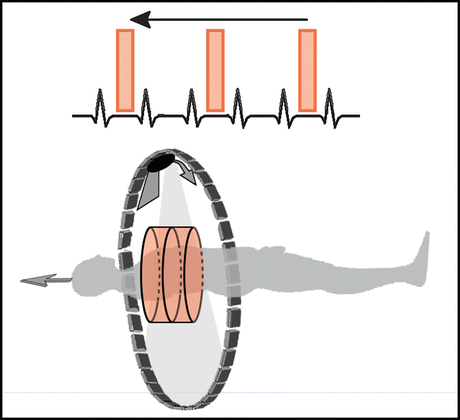
Fig. 5
Prospective ECG triggering: The scan is obtained with a sequential CT during a predetermined phase of the cardiac cycle, according to the ECG
The most important advantage of this technique is a lower radiation exposure (absorbed dose values between 2.1 and 9.2 mSv) [16] compared with the retrospective ECG gating, but it has even a lower temporal resolution (200–250 ms) [15], requiring an HBR necessarily lower than 65 bpm, and it is also more subject to artifacts. Recent studies [17, 18] have shown that prospective ECG-triggering CTA are similar in quality to those obtained with retrospective ECG gating with a lesser dose of radiations (about 76.5 %) [17].
The step-and-shoot technology with dual-source CT in patients with less than 70 bpm allows to obtain diagnostic images in more than 97 % of the cases reducing the amount of dose given to the patient compared to retrospective ECG gating analysis [19].
With the introduction in clinical practice of the dual-source CT scanner, temporal resolution has been further improved, up to 75 ms, allowing a new acquisition mode, known as “flash mode”: this method, thanks to a high pitch (>3.4) and large detector coverage, consents to acquire a heart scan during a single heart beat in a quarter of second with very low radiation doses (<1 mSv) [11].
The examinations can be reconstructed in two methods:
Partial scan reconstruction: With this modality, used either for retrospective ECG gating and prospective ECG triggering acquisition modes, the data used for the reconstruction derived from a predefined R-R interval of a single heart cycle [15].
If the CT scanner uses filtered backprojection reconstruction algorithms, the B46f kernel is the most used in clinical practice; the latest CT scanners use iterative image reconstruction algorithms with a substantial reduction in radiation doses (see below).
Once the exam has been correctly executed, the radiologist using different post-processing techniques analyses it, such as MIP, or volume rendering, which is useful in particular for clinicians to have a simple and anatomic view of the coronaries [20]. Curved multiplanar reformation is widely used because it allows to “stretch” the coronaries, in order to follow the entire course of the vessels and to depict lumen and wall abnormalities [20].
A correct report includes [5] all the information seen above (Table 3).
Table 3
Elements of a correct report
Report data |
|---|
Clinical question for the examination |
Use of drugs before the examination (β-blockers, nitroglycerin, benzodiazepine) |
Acquisition mode (retrospective ECG gating, prospective ECG triggering …) |
IV contrast medium bolus information (type, quantity, administration modality) |
Imaging findings, indicating: • Dominance of the coronary circle (right, left or codominance) • Coronary findings (including malignant and non-malignant anomalies), describing type, location and extension of lesions • Other cardiac findings • Non-cardiac findings |
Before talking about the principal CTA indication and principal image findings in CADs, it is important remembering that nowadays there are many dose reduction strategies which allow to obtain very good examinations with lower radiation doses than in the past; in fact, with the prospective-ECG-triggering and flash acquisition mode seen before we can obtain examination at low doses using lower peak kilovoltage values (100 kV are sufficient if the patient has a BMI <25 kg/m2, and 80 if the BMI is <20 kg/m2) [11]; another way to reduce radiation dose is the use of the iterative image reconstruction algorithms, which allows to obtain CTA with lower radiation doses, lower noise and better quality despite the traditional backprojection algorithms [11, 21–23].
In the pediatric population in order to obtain a diagnostic examination sedation could be required if the little patients are not collaborative with different approaches (midazolam 0.1 mg/kg/dose with the anesthesiologic assistance) [24]. Sedation and breath hold would not be required in newborn and quiet babies, especially if modern faster scanners are used. It is of course important giving the lowest radiation of dose “as low as reasonably achieved” (“ALARA”) to the patient, adopting several strategies, like increasing the pitch value, reducing the volume coverage and tube kV (80 kV with a weight less than 25 kg) and adjusting the value of mAs to the weight of the patient [24]. It is moreover preferable to use the ECG-gated acquisition to limit the motion artifacts, and if available, prospective ECG triggering or the flash acquisition mode than retrospective ECG gating mode.
CTA Indications
Like other radiologic examinations that involve X-rays, CTA can be executed if there is a favourable risk-benefit ratio for the patient, and this risk has to be evaluated individually for every single patient [3]. CTA is indicated for symptomatic patients that on the bases of age, sex, symptoms and previous non-radiologic examinations have a low to intermediate probability of significant CAD, in particular symptomatic young patients with no ECG changes and serial enzymes negative, with an ECG uninterpretable, unable to exercise or stress test, with uninterpretable or equivocal stress test, or with suspected non-aterosclerotic coronary anomalies (such as anatomic anomalies or aneurisms) [3, 25].
A detailed meta-analysis of different studies have confirmed the correlation between CAD severity detected on CTA examinations and increased risk of related heart adverse events: the absence of CAD is related with a low risk of adverse cardiac events, whereas a non-obstructive or obstructive CAD is associated with an increase of the likelihood of adverse cardiac events [26]. Another study [27] confirms these data and extends the outcome over the 5 years.
Other indications are the evaluation of stents and CABG before the execution of a CA.
Thanks to technical improvements and introduction of modern CT scanners (single-source 64-slice or more and dual source) in clinical practice and in Emergency Departments (ED), another emerging indication for CTA is the evaluation of acute chest pain in ED (not as a replacement examination, but done besides clinical evaluation, ECG and serum cardiac enzyme) [11, 14]. In fact, the so-called triple rule out allows to evaluate at the same time, and eventually exclude (or confirm), three important clinical conditions [14]:
Coronary thrombosis
Pulmonary embolism
Thoracic aortic dissection
Currently there is still no indication as screening examination for asymptomatic patients because of the high radiation exposure [3]; thanks to the technological improvements for dose reduction such as flash acquisition mode (see above [11]) we can even imagine that in few years CTA will become a screening examination for CAD.
Dual-source CTA is a useful examination in the pediatric population, in particular in those with a high likelihood of coronary artery anomalies [28], and when associated with ECG-triggered prospective spiral acquisition with high pitch, low tube current tube and iterative reconstruction method [29].
Principal CADs and Imaging Findings
Atherosclerosis is the principal coronary disease, but it is not the only one; in fact, as we have seen before, other pathologic conditions that have been evaluated are the anatomic malignant and non-malignant variants, aneurisms and fistulas and the evaluation of stents and bypass grafts.
Coronary Anatomic Anomalies
Coronary anatomic anomalies are inborn anomalies that occur in 1 % of the general population [30], even if necropsies show lower incidence (0.3 %) [30]. Moreover, it is known that sometimes patients with congenital heart diseases have one or more associated coronary artery anomalies [31]. There are many anatomic variants and could manifest in young patients with symptoms like atypical chest pain shortness of breath or acute myocardial infarction [32], but most of that have no clinical outcome [5]; thereby, some of them are malignant and have to be recognised and described.
Some of these anomalies could be not recognized with the traditional CA [33]. CTA is the best examination available in the study of these clinical conditions [32] because of its intrinsic ability in the global morphologic study of the heart and vessels.
Malignant coronary anomalies (Fig. 6) manifest clinically with chest pain, dyspnoea, syncope, myocardial infarction, ventricular fibrillation or sudden death, rarely with reproducible effort angina [30]. There are many different malignant variants, and Bland-White-Garland syndrome [34] is one of them: even known as ALCAPA syndrome (anomalous origin of the left coronary artery from the pulmonary artery), and consists of the anomalous origin of the left coronary artery (or, sometimes, of the right coronary artery, ARCAPA, which is a more favourable condition [32]) from the pulmonary artery. The incidence is of 1/300,000 live birth, and without therapy the rate of death is 90 % during the first year of life [32]. If this condition is not corrected, and a compensatory and ectasic circle develops between RCA and left coronary circle as compensatory mechanisms, the patient survives and can develop early signs of pulmonary hypertension and of heart failure [32].
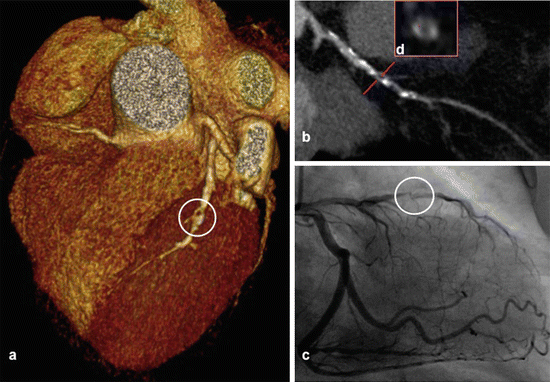

Fig. 6
Volume-rendering reconstruction demonstrates a malignant coronary anomaly with inter-arterial course of the right coronary artery (arrow) that origins from the left sinus of Valsalva
Coronary artery originating from the opposite sinus of Valsalva is another malignant anomaly: in this case, the acute angle formed at the origin of the vessel could be responsible of transitory ischemia during the systole [5].
Coronary artery with origin from the opposite or non-coronary sinus (ACAOS) associated with an interarterial course between the aortic root and common trunk of pulmonary artery is often found in young athletes and militaries, and requires surgical treatment for the high risk of sudden death [5]. The pathophysiological mechanisms of sudden death are still unclear, but it could be explained by a reduced blood flow due to the acute angle which often characterised the emergency of the coronary artery associated with the intramural course and the compression of the artery every systolic phase [32]. ACAOS associated with prepulmonic, intraseptal and retroaortic course are considered benign [32].
Coronary artery fistulas are discussed below in this chapter.
Another important condition is the “ myocardial bridging ” (Fig. 7), an anomalous course of a coronary tract through myocardium, preceded and succeeded by a normal epicardial course [5]. Myocardial bridging is visualised on CA as a systolic narrowing of an epicardial coronary [35], whereas in CTA it is visualised as a tract of coronary surrounded by myocardial fibres using multiplanar reformation [36]. The significance of this condition is still debated, but it has been seen that atherosclerosis is present with higher frequency in the segment proximal to the tunnelled tract and at this level an endothelial dysfunction is moreover present which could predispose to vasospasm and thrombosis [35]. When radiologists encounter a myocardial bridging they have to report it; the treatment, medical or surgical, will be evaluated by the clinical team.
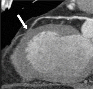

Fig. 7
Long-axis left ventricular multiplanar reconstruction illustrates a myocardial bridging (arrow) of the middle tract of the left anterior descending artery
In the end, it is important to remember that in certain cases we could be unable to find a coronary artery because of an atresia or agenesis, in particular in those cases of single coronary artery. This finding has a prevalence of 0.024/0.066 % in the general population, and could be associated with other congenital heart disorders [37]. It could be totally asymptomatic and find as an incidental finding in the execution of a CTA. The anomaly is classified into three different groups according to the Lipton classification [37]; the prognosis of patients with this pathological condition is not clear: it of course depends on the presence of other heart anomalies associated, but it has even seen that a major cardiac event occurs in 15 % of patient under 40 years [37]. At the moment there are no guidelines on the management of the patient with this condition [37].
Coronary Stenosis: Atherosclerosis and Atherosclerotic Plaques
Pathophysiology and Clinical Manifestation
Coronary atherosclerosis is the most important disease of the coronary arteries. Atherosclerosis is a well-known and described clinical condition related to various factors, including familiarity, tobacco smoke, obesity, hypertension, diabetes, high cholesterol serum levels and reduced physical activity. In particular, it consists of the development of plaque on arterial walls that reduce the vessel lumen, diminishing the blood supply to heart walls.
Stay updated, free articles. Join our Telegram channel

Full access? Get Clinical Tree


