Abstract
Preoperative CT is important in planning functional endoscopic sinus surgery (FESS). An understanding of the sinus anatomy, normal variations, and their clinical implications is important in minimizing unexpected or dangerous complications. This article reviews the normal variations of the paranasal sinuses using the common terminologies found in the FESS literature and highlights their clinical and surgical significance to the reporting radiologist and their surgical colleagues.
Keywords
CT, ENT, FESS, Normal variants, Paranasal sinuses, Sinonasal, Sinusitis
One of the earliest computed tomography (CT) evaluations of the paranasal sinuses was published by Hesselink et al. in 1978 as a two-part series: describing both normal and pathologic anatomy. Early CT studies were performed only in the axial and coronal planes, but with the availability of multiplanar high-resolution CT, more complex imaging anatomy and anatomic variations have been documented. As such, preoperative CT has gained favor among ear, nose, and throat surgeons for the planning of functional endoscopic sinus surgery (FESS). Understanding sinus anatomy and its surgical implications will help the radiologist tailor a surgically relevant report, alert the surgeon to critical anatomic variants, and minimize dangerous complications.
The reported incidence of major complications of FESS ranges between 0.0% and 2.25%. Cerebrospinal fluid (CSF) leak is generally accepted as one of the most common and worrisome complication, associated with significant morbidity. Other complications include bleeding, orbital and optic nerve injury, meningitis, and lacrimal duct injury. It is useful to bear these in mind when reviewing the CT study, as they serve as reminders to highlight important structures.
The Nasal Septum and Nasal Mucosa
The nasal septum is the first structure seen by the endoscopist. Septal deviation is a common deformity, with an incidence of 44.8%, although only 2.1% show obstructive symptoms. It is sometimes associated with a bony spur ( Fig. 2.1 ) and can limit access into the middle meatus or even result in contact point neuralgia as described by Stammberger.

Another common finding is asymmetry of the nasal mucosa and size of the nasal passages. In 1923, Lillie described periodic changes in the nasal mucosa, termed the “nasal cycle.” The thickened nasal mucosa seen on CT is a normal cyclical phenomenon and should not be confused with inflammation. These changes are seen in the turbinates, nasal septum, and ethmoid sinuses, sparing the frontal, maxillary, and sphenoid sinuses. The nasal cycle is believed to be controlled by the suprachiasmatic nucleus in the hypothalamus, and this control decreases with age.
Frontal Sinus Drainage Pathway
The frontal sinus and the frontal sinus drainage pathway (FSDP) are challenging for both radiologists and surgeons, because of their complex and variable pneumatization patterns. A useful imaging landmark is the frontal beak (FB), a bony ridge formed by the superior extension of the frontal process of the maxillary bone ( Fig. 2.2 ). The frontal ostium is found at the level of the FB, which also serves to separate the fontal sinus superiorly from the FSDP inferiorly ( Fig. 2.2 ).

The FSDP is bordered by the most vulnerable parts of the anterior skull base, sandwiched between the lamina papyracea laterally and the thin lateral lamella of the olfactory fossa medially. Here, the bone can measure as little as 0.05 mm and is easily perforated. Its anterior wall is formed by the FB and agger nasi cell (ANC), whereas the posterior wall of the FSDP is the bulla lamella. The FSDP can be further divided into two compartments:
- •
Superior compartment: (“frontal recess”) is formed by a combination of frontal recess cells that can be classified using standardized criteria developed by Lee et al. Its upper border is the frontal ostium.
- •
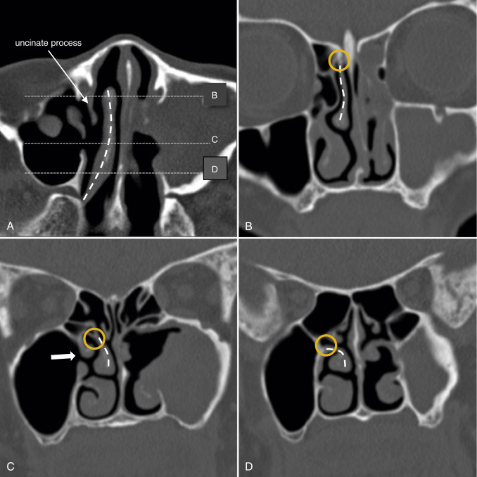
Inferior compartment: (“frontal sinus outflow tract” [FSOT]) is a narrow passageway formed by either the ethmoid infundibulum (38%) or the middle meatus (62%), depending on the variable anatomic attachment of the uncinate process ( Fig. 2.15 ). The patency of the FSOT is also determined by configuration of the surrounding anterior ethmoid cells.
The superior compartment in particular, shows substantial variation in size, shape, and course. This is due to the unpredictable formation of osseous septations and marginating air cells lining the FSDP. It is not uncommon to find more than a single opening of the frontal sinus into the FSDP ( Fig. 2.3 ).

Anterior Ethmoid Cells/Frontal Recess Cells
Multiple named air cells are found surrounding the frontal recess. Anatomically, ANCs and frontal cells (FCs) occur along the anterior wall of the frontal recess, whereas suprabullar cells (SBCs), frontal bullar cells (FBCs), and supraorbital ethmoid cells (SOECs) occur posteriorly along the skull base. SBCs do not extend to the frontal sinus unlike the FBCs. Interfrontal sinus septal cells (IFSSCs) occur along the frontal sinus septum. Apart from the ANC, these cells are collectively known as anterior ethmoid cells (AECs) ( Fig. 2.4 ).

Agger Nasi Cells
The agger nasi cells (Latin for “nasal mound”) are the most anterior ethmoidal cells and extend anteriorly into the lacrimal bone ( Fig. 2.5 ), where they are posteromedial to the lacrimal sac. They are anterior to the anterior attachment of the middle turbinate to the skull base and anteroinferior to the frontal recess, forming the anterior margin of the frontal recess. Because of this intimate relationship, these cells form excellent surgical landmarks and are reliably seen in up to 98.5% of all patients. Opening the ANC usually provides a good view of the frontal recess. The ANC drains into the middle meatus.

Understanding the FSDP is important when evaluating and planning the surgical treatment of frontal sinus disease. At this point, it would also be useful to know that disease within the lateral frontal sinus is difficult to correct endoscopically, often requiring an external approach ( Fig. 2.6 ).

The FSDP is also part of a larger complex known as the ostiomeatal unit (OMU). Its other components comprise the maxillary sinus ostium, ethmoid infundibulum, and hiatus semilunaris ( Fig. 2.7 ). The OMU is the common drainage of the anterior sinuses, and diseased mucosa here impairs ventilation and drainage of the frontal, anterior ethmoid, and maxillary sinuses. FESS is aimed at reestablishing normal ventilation and drainage around the OMU, and the key sinus in FESS is the ethmoid sinus. Its close relationships with the orbit and anterior skull base make these structures vulnerable to trauma during surgery. A good knowledge of the anatomy of the ethmoid bone is essential for understanding the pathologic processes and prevention of surgical complications.

Ethmoid Bone
The word “ethmoid” means “sieve-like” in Greek, largely referring to the porous cribriform plate. It is centrally located within the nasal passage and consists of four parts: the perpendicular plate, the cribriform plate, and two ethmoidal labyrinths ( Fig. 2.16 ). The key ethmoid structures develop as ethmoturbinal attachments (also known as basal lamellae) along the lateral nasal cavity wall. After a series of regression, at least four basal lamellae remain ( Fig. 2.8 ): the uncinate process, ethmoid bulla, middle turbinate, and superior turbinate. Five, if the supreme turbinate is included.

This five basal lamellae concept proposed by Killian is perhaps the best way to understand the ethmoid anatomy, with the four main basal lamellae separating the various draining pathways. The frontal and maxillary sinuses drain into the hiatus semilunaris between the first (ANC and uncinate process) and second (ethmoid bulla) basal lamellae. The ethmoid bulla drains via the superior hiatus semilunaris superior (SHS) and sinus lateralis into the middle meatus between the second and third (middle turbinate) lamellae. The third basal lamella (now referred to in various texts as the “basal lamella”) separates the anterior and posterior ethmoid air cells, whereas the fourth (superior turbinate) lamella separates the posterior and postreme meatus.
Middle Turbinate and Basal Lamella
Owing to their embryology, the middle turbinate and basal lamellae are both part of the same structure (the third basal lamella). It divides the ethmoid cells into the anterior and posterior groups, which is important surgically, as the anterior group drains into the middle meatus, whereas the posterior group drains into the superior meatus.
The middle turbinate has attachments to the skull in all three planes ( Fig. 2.9 ): The anterior vertical part attaches superiorly to the cribriform plate (sagittal plane). The middle oblique part loses its connection to the cribriform plate, attaching to the basal lamella or lamina papyracea (coronal plane). The posterior horizontal part forms the roof of the posterior middle meatus. Here, it attaches to the perpendicular plate of the palatine bone/medial wall of the maxillary sinus (axial plane). This three-dimensional orientation, initially sagittal, then coronal followed by an axial plane, gives the middle turbinate remarkable stability. Resection of the posterior portion may lead to anterior instability.
Middle Turbinate Variants
A concha bullosa is a pneumatized middle turbinate and has a reported prevalence of 34% ( Fig. 2.10 ). A concha bullosa is readily identified on CT, but endoscopic recognition may be difficult. An unremarkable middle turbinate during endoscopy may show extensive pneumatization on CT. Conversely, an endoscopically large middle turbinate may show no pneumatization.

A concha bullosa itself does not necessarily predispose the patient to sinus disease. However, it may obstruct the ethmoid infundibulum. In addition, sinusitis, polyps, and mucoceles may affect the concha bullosa itself. A concha bullosa can be marsupialized, crushed, or excised, but care must be taken to avoid damaging the attachment of the middle turbinate to the skull base. A fracture at this site may lead to an unstable middle turbinate or CSF leakage. A concha bullosa may also occur in the superior turbinate, although this is much less often seen ( Fig. 2.11 ).

Normally, the convexity of the middle turbinate is directed medially. However, in 26% of cases the convexity is directed laterally ( Fig. 2.12 ). This condition is termed paradoxical middle turbinate and is usually of no clinical significance. Turbinate sinus refers to the exaggerated curvature of the middle turbinate, forming an internal concavity ( Fig. 2.12 ).

Related posts:
 Imaging of the Postoperative Middle Ear, Mastoid, and Internal Auditory Canal
 Imaging of the Facial Nerve
 Neurointerventional Radiology for Skull Base Lesions
 Jugular Foramen
 Skull Base Bone Lesions I: Imaging Technique, Developmental and Diffuse Bone Lesions
 Skull Base Bone Lesions II: Benign and Malignant Tumors
Stay updated, free articles. Join our Telegram channel

Full access? Get Clinical Tree


