38 Imaging of urinary tract calculi

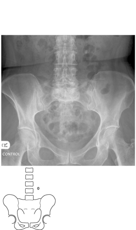
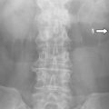
Fig. 38.1 This radiograph demonstrates an opacity to the left of the L3/4 disc space which represents a stone.
Background
Ninety per cent of urinary tract calculi are radio-opaque on a plain radiograph. There are several strategies for imaging suspected urinary tract calculi, which are described below.
Renal colic is a very common presentation to the on-call doctor. Infection proximal to obstruction is an emergency. Remember also that abdominal aortic aneurysms can present similarly.
Clinical features
Symptoms
There is acute loin pain, radiating to the groin. The onset of pain is usually sudden. Nausea and vomiting may also be a feature. The pain is visceral, so the patient is restless, and it is often the worst pain the patient has experienced.
Patients may have a prior history of urinary calculi.
It is important to ascertain whether the patient has signs and symptoms of infection such as pyrexia/rigors (as an obstructed infected kidney is an emergency) and whether they have a single kidney. Obstruction by a stone threatening the integrity of a single kidney is also an emergency which should be addressed immediately.
Differential diagnosis
This is very wide and includes many causes of abdominal pain:
Radiological features
There are three methods for imaging suspected urinary tract calculi: plain radiographs, IVUs and unenhanced CT.
Plain radiographs
The traditional first line was to obtain a plain radiograph, centred on the urinary tract – a ‘KUB’ film (kidneys, ureters, bladder).
It is still useful to be able to assess such a film for ureteric calculi, as you may also detect a ureteric calculus in a patient presenting on the surgical ‘take’ with abdominal pain. But this is now not the first-line investigation if stones are suspected.
A knowledge of the course of the ureter is important for proper assessment of a plain film of the abdomen (see Chapter 28).
Phleboliths – small calcifications in the pelvic small vessels – can be confused with stones but they are generally completely round, and have lucent centres.

Stay updated, free articles. Join our Telegram channel

Full access? Get Clinical Tree

