, Maria Custódia Machado Ribeiro2 and Bruno Beber Machado3
(1)
Hospital da Criança de Brasília – Jose Alencar, Clínica Vila Rica, Brazília, Brazil
(2)
Hospital da Criança de Brasília – Jose Alencar, Hospital de Base do Distrito Federal, Brasília, Brazil
(3)
Clínica Radiológica Med Imagem, Unimed Sul Capixaba, Santa Casa de Misericordia de, Cachoeiro de Itapemirim, Brazil
Abstract
Musculoskeletal infection in children often poses a diagnostic challenge, so much greater as the younger the patient is. Although joint aspiration remains indispensable for definitive diagnosis, imaging is a valuable tool in the workup. No matter the etiologic agent, early and accurate definition of the infectious origin of the arthritis is paramount in order to minimize structural damage and to avoid complications and long-term sequelae. This chapter will emphasize the articular component of musculoskeletal infections as well as related diseases that should be considered in the differential diagnosis, such as transient synovitis and chronic recurrent multifocal osteomyelitis. Spinal infection will be addressed in Chap. 11.
5.1 Introduction
Musculoskeletal infection in children often poses a diagnostic challenge, so much greater as the younger the patient is. Although joint aspiration remains indispensable for definitive diagnosis, imaging is a valuable tool in the workup. No matter the etiologic agent, early and accurate definition of the infectious origin of the arthritis is paramount in order to minimize structural damage and to avoid complications and long-term sequelae. This chapter will emphasize the articular component of musculoskeletal infections as well as related diseases that should be considered in the differential diagnosis, such as transient synovitis and chronic recurrent multifocal osteomyelitis. Spinal infection will be addressed in Chap. 11.
5.2 Pyogenic Arthritis
The term pyogenic arthritis (PA) is used to describe bacterial joint infection, which accounts for approximately 6 % of all childhood arthritides. PA is most often a monoarthritis, more frequent in males with less than 3 years of age. The joints of the lower extremities are involved in approximately 75 % of the patients, mostly the hips and the knees; ankles, elbows, and shoulders are other important sites of disease. Even though PA is usually secondary to hematogenous spread, in children younger than 18 months, the process may begin in a metaphyseal focus that disseminates via transphyseal vessels. These vessels involute in older children, in whom the growth plate acts as a barrier for dissemination. Less common ways of contamination include direct inoculation (open wounds, joint punctures, surgery) or dissemination from a contiguous focus in the adjacent soft tissues.
Time is a critical prognostic factor in PA, as delayed institution of treatment leads to irreversible joint destruction and permanent sequelae. The acute inflammatory response related to bacterial infection causes quick cartilaginous destruction, and increase in the intracapsular pressure – due to synovial hypertrophy and purulent effusion – may lead to joint dislocation and epiphyseal ischemia, mostly in septic hips. Definitive diagnosis relies on joint aspiration and analysis of the synovial fluid, which must be performed as soon as PA is suspected. Imaging plays a secondary role, given that none of the available methods is able to distinguish infectious from noninfectious arthritides and a normal imaging study does not rule out articular infection.
Radiographs must be obtained in all patients, even though this imaging modality is quite insensitive: irreversible joint damage is usually present by the time that radiographic abnormalities become evident (Fig. 5.1). Findings related to the early stages of articular infection are fairly nonspecific, including joint effusion, widening of the joint space, and soft-tissue swelling (Fig. 5.2). Narrowing of the joint space appears quickly, as well as epiphyseal osteopenia (less pronounced than that found in tuberculous arthritis) and peripheral erosions (Figs. 5.3 and 5.4). Despite the fact that the knee is the most affected joint, PA is usually more severe in the hips (Fig. 5.5). Polyarticular PA occurs in less than 10 % of all cases (Fig. 5.6).
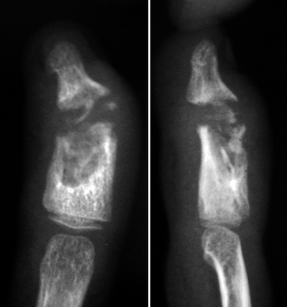
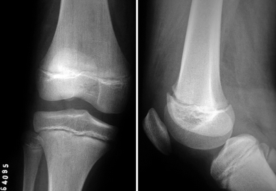
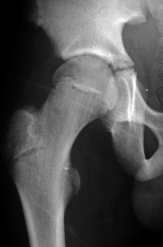
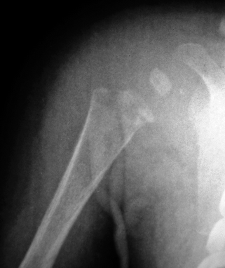
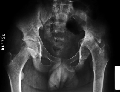
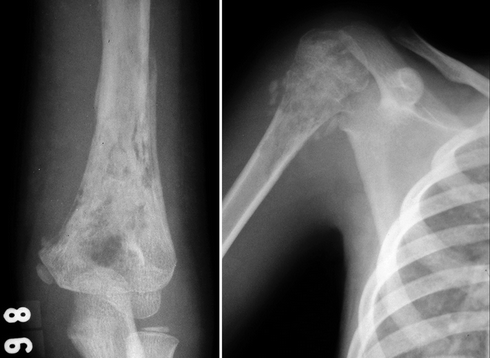
Fig. 5.1
Radiographs of the index finger of the right hand of a 6-year-old female who sustained an open fracture of the middle phalanx 6 months before, presenting spontaneous pus drainage from a fistulous tract and marked soft-tissue swelling. There are signs of chronic osteomyelitis of the middle phalanx, with extensive osteolysis and bone sequestra in the medullary cavity and in the distal interphalangeal joint. Destruction of corresponding joint surfaces due to PA is also evident, with deviation of the distal phalanx
Fig. 5.2
A 9-year-old female with acute osteomyelitis of the right distal femur and secondary contamination of the joint cavity of the knee. There are no osseous abnormalities on radiographs, although swelling of periarticular soft tissues can be seen, mostly suprapatellar and more evident on the lateral view. There is also loss of definition of periarticular fat planes and subtle widening of the joint space. Nonetheless, these are nonspecific radiographic findings
Fig. 5.3
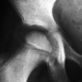
Anteroposterior view of the right hip of a child with PA whose onset occurred less than 1 month earlier. There is osteoporosis of the proximal femur and narrowing of the coxofemoral joint space, as well as irregularity of the acetabulum. Such findings represent advanced cartilaginous destruction
Fig. 5.4
Radiograph of the proximal portion of the right arm of a young child with bacterial osteomyelitis and secondary contamination of the shoulder joint. There are lytic lesions associated with cortical destruction in the proximal metaphysis, as well as marked swelling of periarticular soft tissues and widening of the proximal humeral physis
Fig. 5.5
Pyogenic arthritis of the left hip with poor evolution. There is marked bone destruction, with almost complete resorption of the left proximal femoral epiphysis and bone remodeling of the corresponding acetabulum; acetabular protrusion is also evident, with striking acetabular incongruity
Fig. 5.6
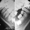
Radiographs of the left elbow (first image) and of the right shoulder (second image) of a child with multifocal bacterial osteomyelitis and secondary articular contamination. There is metaphyseal osteomyelitis in the left distal and in the right proximal humeri, with permeative osteolysis, periosteal reaction, soft-tissue swelling, and epiphyseal involvement, the latter more evident in the shoulder. Osteomyelitis of the right femur and pyogenic arthritis of the homolateral hip were also present (not shown)
Ultrasonography (US) is very useful for early detection of joint effusion and synovitis, being also very appropriate for image-guided joint aspiration. Common findings include joint effusion (often with suspended debris) and hyperemia of the inflamed synovium on Doppler studies, but they are not specific (Fig. 5.7). US is also able to demonstrate erosions (especially large ones) and dissemination of the infection to adjacent bursae and tendon sheaths.
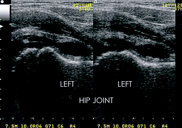
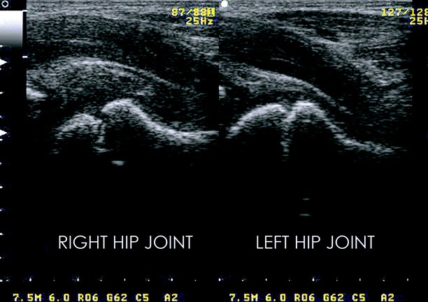
Fig. 5.7
US of the hips of two distinct children, both with pyogenic arthritis. The upper images show longitudinal scans of the left hip joint demonstrating joint effusion associated with irregular synovial thickening. In the lower row, there is a heterogeneous material filling the joint cavity of the right hip, representing synovial thickening and joint effusion with suspended debris. The proximal epiphysis of the right femur is irregular and of reduced size, notably if compared to the contralateral one (Courtesy of Dr. Telma Sakuno, Hospital Universitario da UFSC, Florianopolis, Brazil)
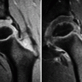
Magnetic resonance imaging (MRI) is capable to identify the earliest changes related to PA, allowing for accurate assessment of disease extent and helping in preoperative planning. It is highly sensitive for joint effusion (which may be heterogeneous) and synovitis, mostly when intravenous contrast is administered, as there is enhancement of the synovium and of the inflamed tissues (Figs. 5.8, 5.9, 5.10, 5.11, 5.12, 5.13, and 5.14). Erosions and destructive changes of bone and cartilage are also clearly seen (Figs. 5.9, 5.10, 5.11, and 5.12), as well as dissemination of the infectious process to nearby bursae and tendon sheaths (Fig. 5.14). Subchondral bone marrow edema is common and not necessarily indicative of osteomyelitis, as it may be merely related to reactive (noninfectious) osteitis. Osteomyelitis is more likely if the edematous areas extend far beyond the subchondral bone, notably if there is prominent low signal intensity on T1-weighted images (T1-WI) (Figs. 5.8, 5.12, 5.13, and 5.14). Cortical discontinuity, periosteal reaction, intraosseous abscesses, and soft-tissue collections are typical of osteomyelitis (Figs. 5.9, 5.12, 5.13, and 5.14).
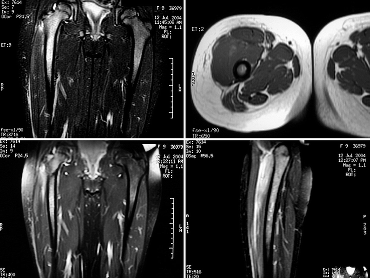
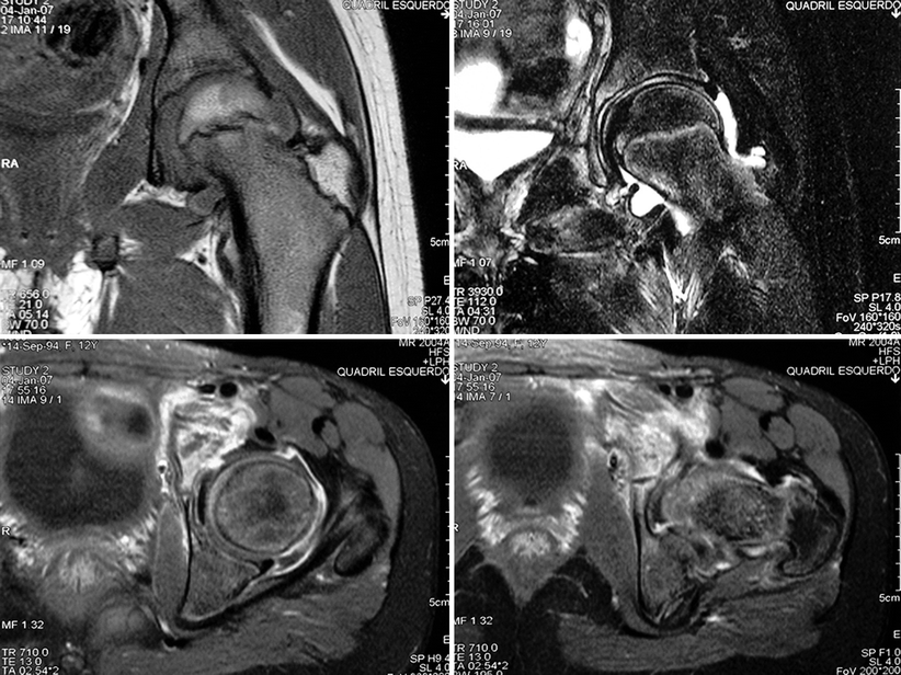
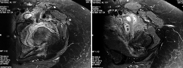
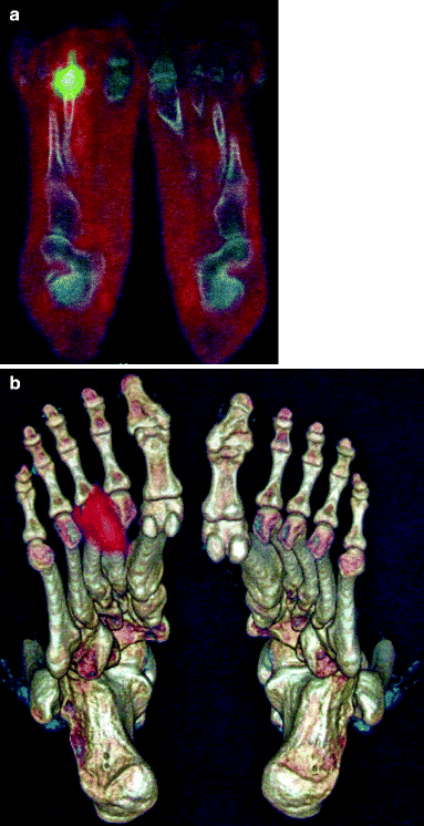
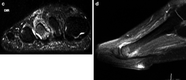
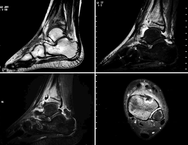
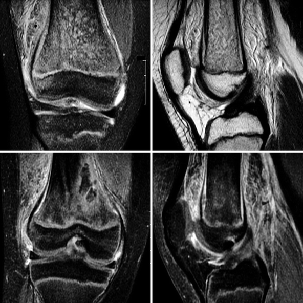
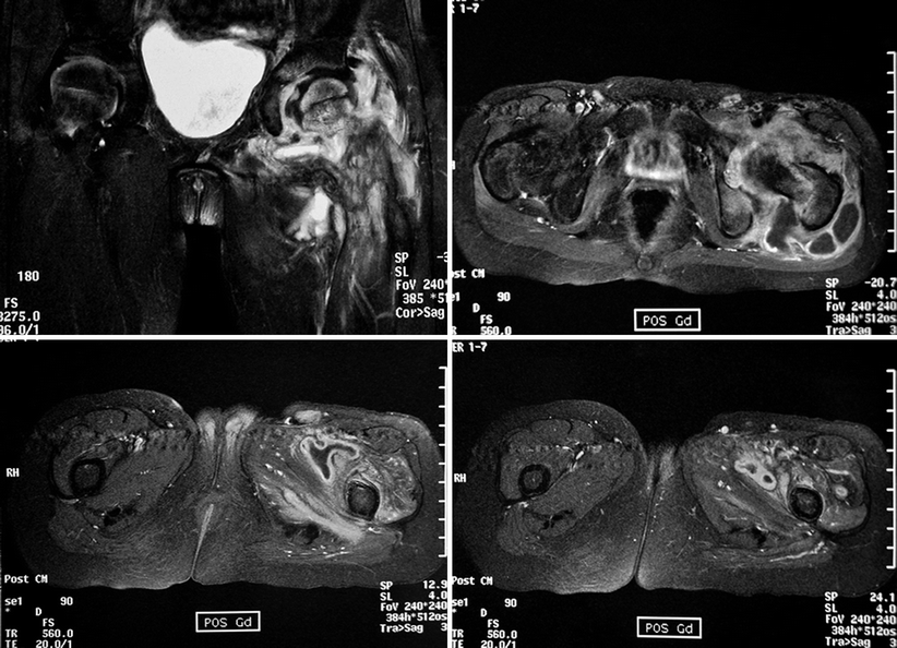
Fig. 5.8
Coronal STIR image (upper-left image), transverse T1-WI (upper-right image), and post-contrast fat sat T1-WI in the coronal and sagittal planes (lower images) of the hips and thighs of a 9-year-old female with right-sided septic coxofemoral arthritis. MRI is very suitable to demonstrate joint effusion in the right hip and bone marrow edema pattern in the ipsilateral femur, which extends far beyond the subchondral bone, reaching the mid-diaphysis, indicative of associated osteomyelitis. There is post-gadolinium enhancement of the edematous bone and of the proximal quadriceps
Fig. 5.9
Coronal T1-WI and fat sat T2-WI (upper row) of the left hip of a 12-year-old adolescent with PA reveal joint effusion and cartilaginous thinning, as well as edematous changes of the soft tissues of the proximal thigh and of the subchondral bone marrow of the acetabulum. Transverse post-contrast fat sat T1-WI (lower row) disclose intense enhancement of the thickened synovium and extensive destruction of the anterosuperior acetabulum, with cortical discontinuity and infiltration of the adjacent soft tissues; contrast enhancement is also seen in the edematous tissues. This appearance may resemble those seen in aggressive bone tumors, like Ewing’s sarcoma
Fig. 5.10
Transverse post-gadolinium fat sat T1-WI of the left hip of a 13-year-old male with PA. Heterogeneous joint effusion, marked synovial thickening, and infected collections in the soft tissues of the proximal thigh can be seen, with post-contrast enhancement. The femoral head is deformed, and its appearance is suggestive of slippage of the proximal femoral epiphysis as a complication of septic arthritis
Fig. 5.11
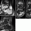
Female adolescent with chronic, low-grade infection in the right forefoot. In (a) and (b), PET-CT demonstrates intense enhancement adjacent to the third metatarsophalangeal joint, initially interpreted as an infection of the periarticular soft tissues with secondary involvement of the contiguous bone. MRI performed 3 months later (coronal STIR image c and sagittal post-gadolinium fat sat T1-WI d) demonstrates that the infection was primarily articular, with joint effusion, synovitis, and erosions in the head of the third metatarsal, as well as edematous changes of the bone and of the adjacent soft tissues
Fig. 5.12
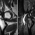
MRI of the left ankle of a 12-year-old male with PA. T1-WI (upper-left image) and STIR image (upper-right image) evidence tibiotalar joint effusion, metaphyseal osteomyelitis of the distal tibia, and a small juxtaphyseal intraosseous abscess, anteriorly situated. Post-gadolinium fat sat T1-WI (lower images) demonstrate the intraosseous abscess with its hypointense content and peripheral enhancement draining to the joint cavity through a cortical break. Synovial enhancement is also present, distinguishing the thickened synovium from the joint effusion
Fig. 5.13
MRI of the same patient of Fig. 5.2, performed on the same day (upper row, coronal fat sat T2-WI and sagittal T2-WI; lower row, coronal and sagittal post-gadolinium fat sat T1-WI). There is diffuse infiltration of the bone marrow of the distal femur, with post-gadolinium enhancement, as well as subperiosteal abscesses, extensive edema of the periarticular soft tissues, and contamination of the joint cavity, with joint effusion and synovitis
Fig. 5.14
MRI of the left hip of a child with PA (upper-left image, coronal fat sat T2-WI; remaining images, transverse post-gadolinium fat sat T1-WI). There is a large and heterogeneous joint effusion on fat sat T2-WI, with extensive bone marrow edema pattern in the proximal femur and acetabulum and edematous changes in the adjacent soft tissues. Post-gadolinium images display a large, complex soft-tissue abscess surrounding the hip and extending to nearby bursae, with thick and irregular enhancement, as well as adjacent myositis
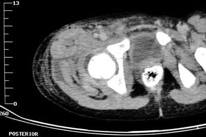
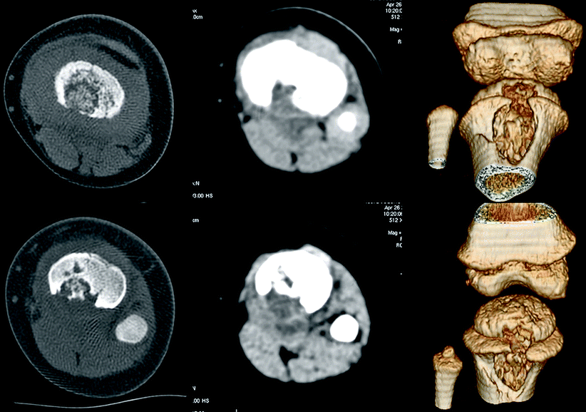
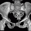
Computed tomography (CT) has restricted usefulness in the assessment of PA in pediatric patients (see Chap. 1). It is usually reserved for selected cases, most commonly used as an alternative (or an adjunct) to MRI for osseous assessment or to evaluate anatomically complex joints (Figs. 5.15, 5.16, 5.17, 5.18, and 5.19); the use of intravenous contrast is formally recommended. If there is associated osteomyelitis, CT is very helpful to demonstrate bone destruction and the presence of sequestra (Figs. 5.16, 5.17, and 5.19). Post-contrast enhancement is more evident in the thickened synovium (Fig. 5.19). CT is the best imaging method to detect gas bubbles in the infected tissues, which are indicative of the bacterial nature of the infection (Fig. 5.15).
 Legg-Calvé-Perthes Disease
 Peculiar Aspects of the Anatomy and Development of the Growing Skeleton
 Musculoskeletal Disorders Related to Pediatric Hematologic Diseases
 Sports-Related Musculoskeletal Lesions in Pediatric Patients
 Juvenile Idiopathic Arthritis
 Juvenile Spondyloarthropathies and Pediatric Collagen Vascular Disorders


Fig. 5.15
A 9-year-old male with PA of the right hip and septicemia. CT scan reveals joint effusion, marked swelling of periarticular soft tissues, densification of the subcutaneous fat, and juxta-articular abscesses. Gas bubbles can be seen in the collections, indicative of the bacterial nature of the infection (Courtesy of Dr. Arthemizio Rocha, Hospital Santa Marta, Taguatinga, Brazil)
Related posts:
Legg-Calvé-Perthes Disease
Peculiar Aspects of the Anatomy and Development of the Growing Skeleton
Musculoskeletal Disorders Related to Pediatric Hematologic Diseases
Sports-Related Musculoskeletal Lesions in Pediatric Patients
Juvenile Idiopathic Arthritis
Juvenile Spondyloarthropathies and Pediatric Collagen Vascular Disorders
Stay updated, free articles. Join our Telegram channel
Full access? Get Clinical Tree
