Abstract
This chapter provides information, images, and interpretative criteria related to nuclear medicine scanning of suspected infection and inflammation. There is discussion of technetium-99m, indium-111 leukocytes, and the use of gallium-67 and fluorine-18 fluorodeoxyglucose.
Keywords
infection, inflammation, labeled leukocytes, gallium-67, FDG, sarcoid
Chapter Outline
Fluorine-18 Fluorodeoxyglucose PET/CT Imaging
A variety of nuclear medicine imaging techniques provide effective methods for the detection and assessment of both clinically apparent and occult infectious and inflammatory conditions. Rather than representing organ-specific techniques, these procedures use radiopharmaceuticals that localize preferentially in inflamed or infected tissue in any location in the body. The available radiopharmaceuticals exhibit varying degrees of nonspecificity and are best used with meticulous clinical correlation. The particular effectiveness of each of the radiopharmaceuticals for infection imaging often depends on the clinical setting and the specific part of the body under scrutiny. Selection of the proper imaging agent is critical to the success of the procedure ( Table 12.1 ). The commonly used agents include the following:
- •
Radiolabeled leukocytes
- •
Indium-111 ( 111 In) leukocytes
- •
Technetium-99m ( 99m Tc) leukocytes
- •
- •
Gallium-67 ( 67 Ga) citrate
- •
Fluorine-18 fluorodeoxyglucose ( 18 F-FDG)
| Radiopharmaceutical and Administered Activity | Time of Imaging | Advantages | Disadvantages | Common Uses |
|---|---|---|---|---|
| 111 In white blood cells 300–500 µCi (11.1–18.5 MBq) | 12–24 hr | No interfering bowel/renal activity Delayed imaging possible Simultaneous 99m Tc-sulfur colloid or 99m Tc-diphosphonate bone imaging possible | Less sensitivity for nonbacterial and nonpyogenic infections 111 In label not ideal for imaging Complex preparation Low sensitivity for discitis | Bacterial infections Indolent inflammatory conditions (e.g., prosthetic joint infections) Abdominal infections Prosthetic vascular graft infections Brain abscess Complicated osteomyelitis Extremity infections (e.g., diabetic foot) Renal infections FUO: acute phase |
| 99m Tc-white blood cells 5–10 mCi (185–370 MBq) | 0.5–4.0 hr | Early imaging Excellent early sensitivity 99m Tc label ideal for imaging | Less sensitivity for nonbacterial and nonpyogenic infections Delayed imaging not ideal Early renal activity Bowel activity after 1–2 hours Complex preparation Low sensitivity for discitis | Bacterial infections Acute inflammatory conditions (e.g., inflammatory bowel disease) Complicated osteomyelitis Extremity infections: diabetic foot Osteomyelitis Prosthetic vascular graft infections |
| 67 Ga-citrate 5–10 mCi (185–370 MBq) | 24–48 hr | A variety of infections detected, including opportunistic | Interfering bowel and renal activity Delayed imaging necessary 67 Ga not ideal for imaging | Immunocompromised patients Chronic infections Discitis/spinal osteomyelitis FUO: chronic phase |
| 18 F-FDG PET/CT 5-10 mCi (185-370 MBq) | 1–2 hr | Excellent spatial localization Very sensitive | Not currently FDA approved for infections Nonspecific; also localizes in tumors | Sarcoidosis Peripheral bone osteomyelitis (not diabetic or postoperative) Non-postoperative spinal infection FUO Metastatic infection Primary vasculitis |
Radiolabeled Leukocytes
Leukocyte imaging using in vitro labeling with 111 In-oxine or 99m Tc-exametazime is currently the nuclear medicine gold standard for diagnosing most infections in patients who are not immunocompromised. Because leukocytes can be separated and labeled without significant loss of function, they can be used to image inflammatory processes. Both 111 In-oxine leukocytes and 99m Tc-hexamethylpropyleneamine oxime (HMPAO) leukocytes have been shown to retain their innate function and have demonstrated relatively high sensitivity and specificity for acute infections. However, sensitivity may be somewhat lower for chronic infections. The procedure involves removing some of the patient’s own leukocytes, labeling them, and reinjecting them before scanning. As with any autologous labeled biologic agent, extreme care must be taken to maintain the integrity of the blood sample and to ensure that reinjection of the labeled leukocytes is performed only in the patient from whom the cells were taken. Clinical studies comparing 99m Tc and 111 In-leukocytes have not shown any intrinsic differences in sensitivity for infection when standard 24-hour imaging is performed. However, some notable differences between the two radiopharmaceuticals make one or the other preferable in certain clinical situations.
Mechanism of Localization
Radiolabeled leukocytes are attracted to sites of inflammation, where they are activated by local chemotactic factors and pass from the bloodstream through the vascular endothelium into the soft tissues. The leukocytes then move toward the site of inflammation in a directed migration called chemotaxis . If the inflammation has an infectious cause, the labeled neutrophils phagocytize and destroy any offending bacteria. Gamma camera imaging localizes these accumulations of radiolabeled leukocytes and thus reveals the site of inflammation or infection. Like gallium uptake, radiolabeled leukocyte uptake is not specific for infection and may occur in any inflammatory process that incites a leukocyte response. Occasional uptake in neoplasms may be noted.
Indium-111 Oxine Leukocytes
Labeling Principle
Leukocyte imaging became possible after the development of successful methods for labeling leukocytes with 111 In-oxine. Indium-111 oxine labels all cell types indiscriminately, including platelets and red blood cells. Thus the leukocytes must be isolated from about 50 mL of anticoagulated blood before labeling, commonly through a process called gravity sedimentation , which simply consists of allowing the blood to sit for the time necessary for the red blood cells to settle to the bottom. Any red blood cells remaining in the supernatant may be either lysed by using hypotonic saline or ammonium chloride or removed by centrifugation.
Oxine forms a lipid-soluble complex with 111 In, which passively diffuses through the leukocyte cell membrane. Once this complex becomes intracellular, the 111 In separates from the oxine and binds to cytoplasmic components. The oxine then leaves the cell and is removed by washing the cells. A mixed population of leukocytes is usually labeled, although neutrophils constitute the majority. Labeling efficiencies are on the order of 95%. The patient’s circulating granulocyte count should be at least 3 × 10 6 cells/mL to have enough cells to label.
Minimal manipulation of the leukocytes is essential during the labeling procedure to avoid damage to the cells, which could diminish their viability and thus limit their effectiveness as an imaging agent. Failure to preserve normal physiologic leukocyte function may result in false-negative imaging study results.
Technique
Approximately 300 to 500 µCi (11.1 to 18.5 MBq) of 111 In-oxine–labeled autologous leukocytes are administered intravenously. Care should be taken to avoid excessive agitation of the leukocytes because this may cause clumping, resulting in focal lung accumulation. Although some abscesses can be detected in the first few hours after the administration of labeled leukocytes, most imaging is performed 18 to 24 hours after administration. If the urgency of the clinical setting dictates, 4- to 6-hour images may be useful. A whole-body scan can be performed by using a medium-energy collimator, with gamma camera spot images obtained of specific areas of interest as needed. Generally, both the 173- and 247-keV gamma emissions of 111 In are used. A sample imaging protocol and radiation dosimetry are presented in Appendix E .
Normal Scan
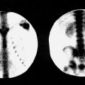
In the first few hours after administration of 111 In-leukocytes, activity is noted in the lungs (likely as a result of leukocyte activation), liver, spleen, and blood pool. The lung and blood pool activity decreases during the first few hours as spleen and liver activity increases. By 18 hours, no lung or blood pool activity is detected, but bone marrow activity is noted ( Fig. 12.1 ).

Twenty-four hours after administration, the 111 In-leukocyte preparation may be found in the liver, spleen, and bone marrow, with the spleen providing the most prominent accumulation, significantly more than that in the liver. No renal or bowel activity is normally present. Damaged leukocytes that remain labeled may provide increased activity in the liver, if slightly damaged, and increased lung activity if severely damaged.
Clinical Applications
General Considerations
Indium-111 leukocytes are taken up nonspecifically at sites of inflammation that incite a leukocytic response regardless of the presence or absence of infection ( Box 12.1 ). The sensitivity (90%) and specificity (90%) are greatest for acute pyogenic infections of less than 2 to 3 weeks’ duration, when leukocytes are still rapidly accumulating. Their effectiveness for detecting more chronic infections is somewhat controversial, although sensitivity with mixed cell populations, which include lymphocytes and chronic inflammatory cells as well as neutrophils, generally appears high (80% to 85%). This may also be because some common bacterial infections may demonstrate significant levels of neutrophil infiltration for months. Labeled leukocytes are of no use in the detection of viral and parasitic infections. Factors that can theoretically reduce leukocyte function, including antibiotics, steroids, chemotherapeutic agents, hemodialysis, hyperalimentation, and hyperglycemia, do not appear to diminish labeled leukocyte sensitivity for detecting infection.
Chest
Common Causes
Adult respiratory distress syndrome
Emphysema
Pleural tubes
Noninfected intravenous lines
Pneumonia
Uncommon Causes
Aspiration
Atelectasis
Cystic fibrosis
Graft infection
Herpes esophagitis
Abdomen
Common Causes
Enteric tubes
Ostomies
Phlegmon
Swallowed leukocytes
Wound infection
Uncommon Causes
Acute enteritis
Bowel infarction
Colitis
Crohn disease
Decubitus ulcer
Gastrointestinal bleeding
Graft infection
Pancreatitis
Transplant (with or without rejection)
Diverticulitis
Acute cholecystitis
Musculoskeletal and Skin Uptake
Common Causes
Intravenous site
Osteomyelitis
Sinusitis
Uncommon Causes
Lumbar puncture site
Rheumatoid arthritis
Septic arthritis
Any Body Part
Abscess
Cellulitis
Wound infection
Hematoma
Infected tumor
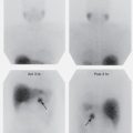
Indium-labeled leukocytes are the preferred radiopharmaceutical for imaging abdominal infection, although in practice, CT scans are usually the initial imaging study ordered for abdominal pain or suspected infection. Because of the lack of normal bowel activity, 111 In-labeled leukocytes have a significant advantage over 99m Tc-labeled leukocytes in diagnosing abdominal abscesses with high sensitivity (85% to 95%). The presence of considerable hepatic and especially splenic activity may hamper the detection of infection in the upper abdomen. Splenic bed abscesses with intense activity may even be confused with a normal spleen ( Fig. 12.2 ), although CT usually resolves this issue. Uncomplicated pancreatitis is usually negative, but septic complications are often imaged successfully.

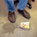
Labeled leukocyte activity in the gastrointestinal tract is nonspecific and may indicate a number of pathologies, including Crohn disease, ulcerative colitis ( Fig. 12.3 ), pseudomembranous colitis, diverticulitis, various gastrointestinal infections, fistulas, ischemic or infarcted bowel, and even vigorous enemas administered before imaging. Increased activity in the bowel, especially the colon, may also be problematic in that activity may be found in the absence of true gastrointestinal disease. False-positive bowel activity is generally caused by swallowing of leukocytes in patients with endotracheal or nasoesophageal tubes, respiratory tract infections, sinusitis, or pharyngitis or in patients with gastrointestinal bleeding of any cause. In general, the more intense the bowel activity compared with liver activity, the more likely it is to indicate a true positive study.

Normal transient physiologic lung activity on early images (1 to 4 hours) severely limits usefulness of 111 In-leukocytes in evaluating pulmonary abnormalities. Increased lung activity is of low specificity at 24 hours as well, because it may occur in numerous infectious and noninfectious processes, including atelectasis, congestive heart failure, pulmonary emboli, aspiration, pneumonia, and adult respiratory distress syndrome. Only one-third of patients showing focal or diffuse uptake in the lungs have infectious causes, although focal uptake demonstrates a slightly better correlation with infection.
Fever of Unknown Origin (Occult Fever)
In a genuine fever of unknown origin (FUO), the spectrum of possible pathology is extensive. The three major categories that account for most FUO are infections, malignancies, and noninfectious inflammatory disease. In occult fevers with a strong suggestion of a pyogenic cause, leukocyte scans may be the study of choice when CT or other anatomic imaging procedures cannot localize the disease. However, there is evidence indicating that positron emission tomography (PET)/CT with 18 F-FDG, although not specific for infection, may be the most efficient method for evaluation of FUO. The value of either 18 F-FDG or 67 Ga scanning in FUO is the ability to detect the spectrum of causative pathologies rather than just infection.
Immunocompromised Patients
Labeled leukocytes have significant limitations for imaging suspected infections in immunocompromised patients. They are not useful in the detection of viral or parasitic infections, demonstrate low specificity in the lungs, and are insensitive for determining active lymph node diseases in these patients. False-negative examinations have been reported in tuberculous and fungal infections. In addition, the study may be technically difficult in severely leukopenic patients. However, 111 In-labeled leukocytes are often useful to evaluate suspected acute pyogenic infections, including sinusitis, bowel infections, and bacterial pneumonias in this setting.
Musculoskeletal Infections
Because labeled leukocytes are taken up by the bone marrow, normal, posttraumatic, or postsurgical variations in marrow distribution may produce confusing foci of increased activity at a site of suspected infection owing to regionally increased marrow uptake. Such false-positive results in the marrow-bearing skeleton can be avoided by performing marrow imaging with 99m Tc-sulfur colloid simultaneously (obtained using the different photopeaks of 99m Tc and 111 In) for comparison with labeled leukocyte distribution. Technetium-99m colloid activity 1 hour after injection is compared with 111 In-leukocyte activity at 24 hours in the area of interest. Criteria for a positive study are (1) spatial incongruence (i.e., leukocyte activity in the absence of sulfur colloid activity) or (2) incongruence of intensity of activity (i.e., leukocyte activity considerably greater than corresponding colloid activity) ( Fig. 12.4 ). Throughout the skeleton, false-positive results may also be produced by nonspecific uptake of labeled leukocytes in recent fractures, heterotopic bone formation, recent radiation therapy, some neoplasms, and noninfectious inflammation, including that caused by gout and rheumatoid arthritis. Simultaneous bone scans may be valuable in leukocyte imaging of the hands and feet to provide anatomic detail needed to separate soft tissue from bony activity.