Inflammatory Arthritis
KEY FACTS
Terminology
Imaging
IMAGING
General Features
Radiographic Findings
 Often normal in early stages of disease
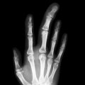
 Soft tissue swelling, juxtaarticular osteopenia, joint space narrowing, bone erosion
 Joint obliteration and ankylosis, particularly of carpus
 Arthritis mutilans and joint deformities (“swan neck,” “boutonniere”) less commonly seen with disease-modifying therapy
MR Findings
 Shows fluid and edematous tissue (synovium, erosions, subchondral marrow, paraarticular soft tissues)
 Effusions and pannus are both hyperintense and may not be readily distinguishable
 Joint space narrowing, erosions, bone marrow edema graded semiquantitatively [outcome measures in rheumatology clinical trials (OMERACT) score]
Ultrasonographic Findings
Imaging Recommendations
 Joints best examined in long axis, tendons in short axis
 Schematic examination of each symptomatic joint
 If multiple joints symptomatic, examine wrists and 3 most symptomatic other joints
 Erosions usually associated with surface cortical irregularity and adjacent pannus
 Use minimal transducer pressure when examining superficial joints, especially for color Doppler imaging
Inflammatory Arthritis


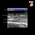
 on the dorsum of the carpus deep to the extensor tendons
  . The distal end of the radius
  , the lunate
  , and the capitate
  are shown.

 on the dorsum of the carpus deep to the extensor tendons
  . The distal end of the radius
  , the lunate
  , and the capitate
  are shown. Hypoechoic synovium is more edematous than hyperechoic synovium.