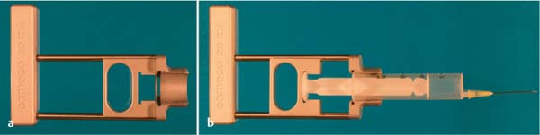
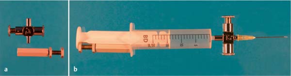
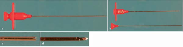
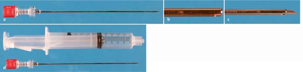
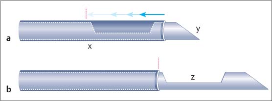
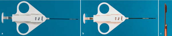
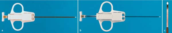
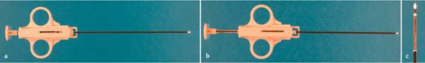
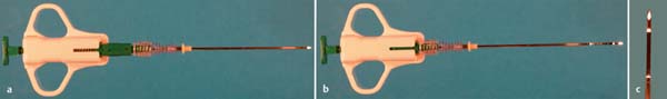
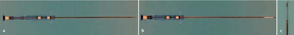
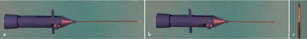
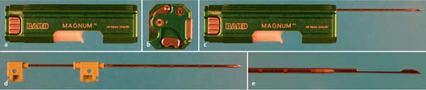
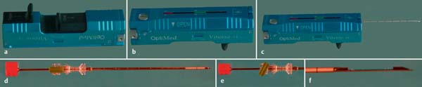
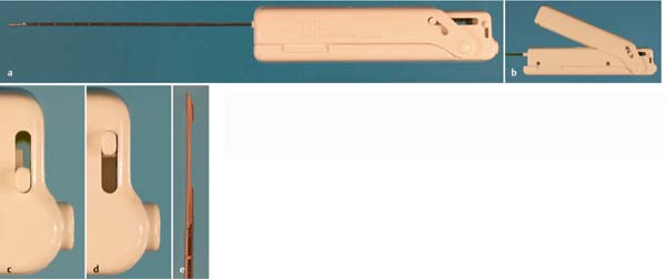
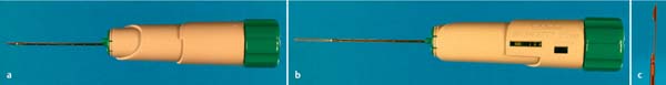
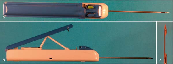
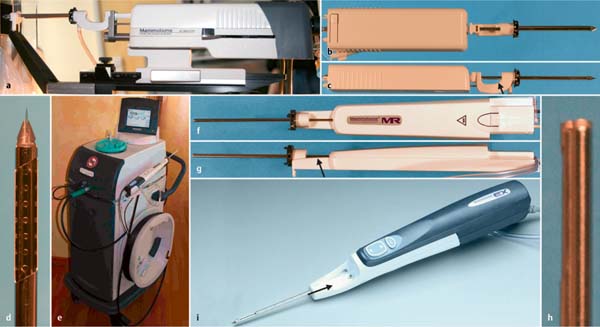
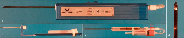
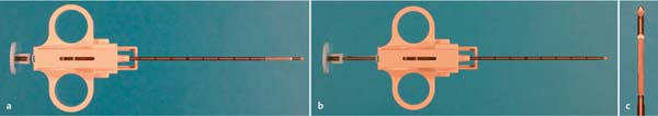
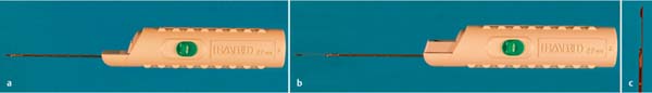
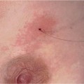
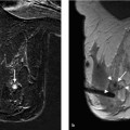
7 Instrumentation The instrumentation used in breast diagnosis includes devices for tissue sample collection and tools for marking lesions to be examined. Special examination equipment and positioning aids facilitate optimal access to the breast. However, some conventional biopsy devices can affect certain key imaging procedures. For example, some biopsy devices produce considerable artifacts in magnetic resonance imaging (MRI); those that do not carry the label “MRI-compatible.” Various techniques are available for specimen collection. Fine needle aspiration biopsy (FNAB) is used for collecting single cells (cytology). Core needle biopsy (CNB) and vacuum-assisted biopsy (VAB) facilitate the collection of tissue samples (histology). Fine needle aspiration (Figs. 7.1, 7.2, 7.3, 7.4, 7.5, 7.6, 7.7, 7.8) uses biopsy needles with outer diameters of 19–26 gauge. Needle tips are available in different point styles. The Chiba needle (Figs. 7.4 and 7.5) has a beveled tip that permits easy puncture of the tissue. As the needle advances, asymmetric tissue displacement at the needle tip causes a shift away from the beveled side. The crown-point tip of the Franseen needle (Fig. 7.6) facilitates the extraction of cells during puncture. When the stylet is introduced, the needle has a symmetric point. Fine needle aspiration is almost exclusively performed under ultrasound (US) guidance. For this reason, it is essential that the examiner be able to guide the syringe and advance the plunger with one hand, while holding the ultrasonic transducer with the other. This is possible with special puncture aids (Figs. 7.1 and 7.2). Fig. 7.1 Cameco handle. Cameco handle without syringe (a), and with cylinder and plunger resting in the corresponding nuts of syringe holder and movable plunger guide (b). The design enables the examiner to determine the needle direction by angulation of the entire holder and, at the same time, to create negative pressure by operating the plunger with the same hand. Fig. 7.2 Binder valve. The Binder valve is used with commonly available syringes. It consists of a valve and a spacer (a). The valve is attached to the Luer nose between puncture needle and syringe cylinder (b). With the valve closed, the syringe plunger is completely pulled back and fixed by the spacer, thus maintaining negative pressure. Once the needle tip has reached the lesion to be examined, suction is produced by pressing the Binder valve. Fig. 7.3 Beveled puncture needle. Puncture needles that are commonly used for collecting blood may also be used for puncturing tissues or cysts. Because these needles do not have a stylet, negative pressure should be created only after penetrating the target lesion. Needle sizes: 19, 20, 21, 22, 23, 24, 25, 26 gauge; lengths: 25, 30, 40, 50, 60, 70, 80, 120 mm. Fig. 7.4 Chiba biopsy needle (Cook Medical, Bloomington, IN). Chiba needle with stylet (a), partially withdrawn stylet (b), beveled needle point (c). Needle sizes: 18, 20, 2-, 22, 23 gauge; lengths: 10, 15, 20 cm. Available also as EchoTip needle designed to enhance needle visibility during ultrasonic imaging. Needle sizes: 18, 20, 21, 22 gauge; lengths: 10, 15, 20, 25 cm. Fig. 7.5 Chiba needle (Bard Biopsy Systems, Tempe, AZ). Chiba needle with stylet (a), Chiba needle with mountable plastic wing and stylet clip for optimal handling (b), beveled needle point (c). Needle sizes: 18, 20, 22, 23 gauge; lengths: 9, 12, 15, 22, 28 cm. Fig. 7.6 Franseen needle (Cook Medical, Bloomington, IN). Franseen needle with stylet (a), needle point with stylet (b), needle point without stylet, showing crown-point tip (c). Needle size: 18 gauge; length: 15 cm. Available also as EchoTip needle designed to enhance needle visibility during ultrasonic imaging. Needle size: 22 gauge; length: 15 cm. Fig. 7.7 Needle for histology/cytology (Bard Biopsy Systems, Tempe, AZ). Histology/cytology needle with stylet (a), needle and stylet separately (b). The histology/cytology needle has no point but a sharpened edge (c). The stylet has a lancet point and extends beyond the needle opening (d). After penetrating the target lesion with the needle point, the stylet is used to create negative pressure for aspiration in the needle by pulling it back. Needle sizes: 18, 20, 21 gauge; lengths: 10, 15, 20 cm. Fig. 7.8 Core biopsy needle (Bard Biopsy Systems, Tempe, AZ). The core biopsy needle (a) has no point but a sharpened edge (b). The stylet is beveled and extends beyond the needle opening (c). The set includes a syringe with a plunger stopper (d) for maintaining the negative pressure in the needle while directing the needle. Needle sizes: 16, 17, 18, 20, 21 gauge; lengths: 5, 10, 15, 20, 28, 40 cm. The aim of performing a CNB is to collect cohesive cell clusters. A typical biopsy needle (Fig. 7.9) is composed of a hollow cannula (x) with a puncture needle inside (y); the puncture needle has at its end a tissue reservoir (notch) (z). First, the puncture needle penetrates the target lesion; then the cannula cuts off the tissue inside the notch, and a specimen of the glandular tissue is recovered with the needle. Characteristic features of a biopsy needle are needle size and needle advance (stroke). There are semi-automatic (Figs. 7.10, 7.11, 7.12, 7.13, 7.14, 7.15, 7.16) and automatic biopsy devices (Figs. 7.17, 7.18, 7.19, 7.20, 7.21, 7.22). The cannula of a semi-automatic biopsy device has a spring mechanism that is tensioned using a plunger on the handle. The puncture needle is advanced into the target lesion by hand, and the cannula then cuts off the tissue by means of spring action. Semi-automatic devices are offered as single-use only. Fig. 7.9 Diagram of a biopsy needle. Biopsy needle before and after biopsy (a), and with the puncture needle advanced (b). Fig. 7.10 Quick-Core biopsy needle (Cook Medical, Bloomington, IN). Quick-Core biopsy needle (a), in the cocked position (b), advanced specimen notch (c). Needle sizes: 16, 18, 20 gauge; lengths: 6, 9, 15 cm; stroke: 10 mm. Needle sizes: 14, 16, 18, 20 gauge; lengths: 6, 9, 15, 20 cm; stroke: 20 mm. Fig. 7.11 Biopsy-Handy semi-automatic biopsy device (SOMATEX Medical Technologies GmbH, Teltow, Germany). Biopsy-Handy biopsy device (a), in the cocked position (b), advanced specimen notch (c). MRI-compatible; needle sizes: 14, 16, 18, 20 gauge; lengths: 10, 15, 20 cm; stroke: 20 mm. Fig. 7.12 Quick-Shot semi-automatic biopsy system (SOMATEX Medical Technologies GmbH, Teltow, Germany). Quick-Shot biopsy device (a), in the cocked position (b), advanced specimen notch (c). Needle sizes: 14, 16, 18, 20 gauge; length: 10 cm; stroke: 15 or 22 mm, optional. Fig. 7.13 Semi-automatic biopsy gun (Invivo Corp., Orlando, FL). Invivo semi-automatic biopsy device (a), in the cocked position (b), advanced specimen notch (c). MRI-compatible; needle size: 14, 16, 18 gauge; lengths: 10, 15 cm; stroke: 20 mm. Fig. 7.14 LUX 2 semi-automatic biopsy device (OptiMed, Ettlingen, Germany). LUX 2 semi-automatic biopsy device (a), with advanced puncture needle (b), notch (c). Needle sizes: 14, 16, 18 gauge; lengths: 10, 16, 20 cm; stroke: 12 or 25 mm, optional. Fig. 7.15 Unicut needle biopsy device (Bard Biopsy Systems, Tempe, AZ). Unicut needle biopsy device (a), with advanced puncture needle (b), notch (c). The system is without a spring mechanism and is shown here only for the sake of completeness. To obtain a tissue specimen, the notch of the puncture needle is first advanced into the target tissue, then the cutting cylinder is manually advanced beyond the notch. Needle sizes: 14, 16, 17, 18 gauge; lengths: 7.5, 11.5, 15, 20 cm; stroke: 20 mm. Fig. 7.16 Autovac disposable full cut biopsy instrument (Bard Biopsy Systems, Tempe, AZ). Autovac single-use biopsy needle (a), in the cocked position (b), needle point (c). This jet needle does not have a notch. The cannula itself acts as the cutting cylinder for obtaining the specimen. Recovering the specimen is occasionally problematic if the tissue is firm, since the tissue sample is not mechanically cut off at the needle point but remains connected with the breast tissue. Detachment is facilitated by rotating the needle after the jet advance or by gentle gyrating movements of the needle tip. Needle sizes: 17, 18, 20, 21 gauge; lengths 10, 15, 20 cm; stroke: 20, 30, or 40 mm, optional. The cannula and puncture needle of an automatic biopsy device are operated with spring mechanisms that need to be tensioned prior to using the device. Here, the needle is placed in front of the target lesion; the sudden advance of the puncture needle and the advance of the cannula occur automatically after pulling the trigger. Reusable automatic devices (Figs. 7.17 and 7.18) are available in addition to single-use devices (Figs. 7.19 and 7.22). A reusable biopsy needle is inserted into a sterilizable needle holder. Different needle sizes and lengths are available. Automatic biopsy devices have a variable stroke. Fig. 7.17 Magnum reusable biopsy system (Bard Biopsy Systems, Tempe, AZ). Magnum automatic hand-held device (a), mechanism for setting needle advance and unlocking the system (b), device with biopsy needle (c), disposable biopsy needle (d), advanced specimen notch (e). Needle sizes: 12, 14, 16, 18, 20 gauge; lengths: 10, 13, 16, 20 cm; stroke: 15 or 22 mm, optional. Fig. 7.18 Vitesse reusable biopsy system (OptiMed, Ettlingen, Germany). Vitesse automatic hand-held device (a, b), device with biopsy needle (c), biopsy needle (d), turning knob for arresting biopsy needle (e), advanced specimen notch (f). If necessary, shipping of a tissue specimen inside the needle is possible by arresting the puncture needle using the turning knob. Needle sizes: 19.5, 18, 16, 14 gauge; lengths: 10, 15, 20, 28 cm; stroke: optional from 10 to 22 mm. Fig. 7.19 TSK disposable automatic biopsy gun (Invivo Corp., Orlando, FL). TSK automatic biopsy device (a), cocking mechanism (b), locked (c), unlocked (d), needle point with notch (e). Needle sizes: 14, 16, 18 gauge; lengths: 10, 15 cm; stroke: 20 mm. Fig. 7.20 Max-Core disposable core biopsy instrument (Bard Biopsy Systems, Tempe, AZ). Max-Core automatic biopsy device (a), in the cocked position (b), specimen notch (c). Needle sizes: 14, 16, 18, 20 gauge; lengths: 10, 16, 20 cm; stroke: 20 cm. Fig. 7.21 Monopty disposable core biopsy instrument (Bard Biopsy Systems, Tempe, AZ). Monopty automatic biopsy device (a), in the cocked position (b), specimen notch (c). Needle sizes: 14, 16, 18, 20 gauge; lengths: 10, 16, 20 cm; stroke: 22 or 11 mm. Fig. 7.22 Celero vacuum-assisted core biopsy device (Suros Surgical Systems, Inc., Indianapolis, IN). Suros Celero spring-loaded, disposable biopsy device (a), lever mechanism for cocking the needle and creating negative pressure (b), needle point with notch (c). This instrument is a hybrid between a CNB needle and a VAB device. By using a vacuum during jet biopsy, the quality of the biopsy specimen is improved. Prior to triggering the needle advance, negative pressure is created at the needle point. This is done to prevent displacement of tissue during jet biopsy and to achieve better filling of the notch. A hinged lever on the handle operates the mechanism of this biopsy device. Two knobs are moved during tissue sampling: first, the spring mechanism is cocked and a vacuum created, then the needle is advanced. Needle size: 12 gauge; length: 12.25 cm; stroke: 25 mm. Unlike other biopsy methods, VAB systems use needles with a lateral aperture into which the tissue is pulled by negative pressure. A cutting cylinder extracts a sample from the tissue. By rotating the aperture, several cubic centimeters of tissue can be removed from around the center of the needle position. Needle sizes vary from 8 to 11 gauge. Fig. 7.23 Mammotome biopsy system (Ethicon Endo-Surgery, Cincinnati, OH). Stereotactic Mammotome (a–e). Mammotome for stereotactic breast biopsy (a), biopsy needle viewed from above (b) and from the side (c), coaxial needle point with tissue acquisition aperture and vacuum holes (d), vacuum pump (e). The Stereotactic Mammotome encloses a spring mechanism to trigger the jet advance. A free-standing vacuum pump is connected to the needle holder by a tubing system. The pump creates negative pressure for pulling tissue through the aperture into the biopsy needle, and also for aspirating blood from the biopsy cavity. The tissue specimen is cut out by a rotating cutter inside the needle. By withdrawing the hollow cutter within the needle, the specimen is moved backward and can be collected at the specimen collection chamber (arrows), while the biopsy needle itself remains in the lesion under examination. This step is repeated with every rotation of the aperture. Needle sizes: 14, 11, 8 gauge; lengths: 8.2 or 9.2 cm. Mammotome magnetic resonance (MR) biopsy system (f–h). For magnetic resonance imaging (MRI-) guided tissue collection, a special hand-held device without jet advance and a special coaxial needle are available. View from above (f), view from the side (g), MRI-compatible coaxial needle with blunt point (h). The vacuum pump remains outside the magnetic field. MRI-compatible; needle sizes: 11 and 8 gauge; lengths: 11.5 and 14.5 cm. Mammotome for ultrasound(US)-guided biopsy (i). Fig. 7.24 Vacora VAB system (Bard Biopsy Systems, Tempe, AZ). Vacora needle holder with the needle inserted (a), sliding carriage for stereotactic biopsy, with fixed coaxial needle (b), single-use needle (c), needle point with notch (d). The Vacora system has a syringe cylinder (c
Fine Needle Aspiration Biopsy


Core Needle Biopsy
Semi-automatic Biopsy
Automatic Biopsy
Vacuum-assisted Biopsy Systems (Figs. 7.23, 7.24, 7.25, 7.26)
![]()
Stay updated, free articles. Join our Telegram channel


Full access? Get Clinical Tree


