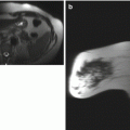
Fig. 5.1
Sagittal MR images of the left breast. (a) T1-weighted fat-saturated image. (b) T2-weighted fat-saturated image. (c) Post-contrast subtraction image. (d) T1-weighted fat-saturated post-contrast image with computer-aided detection (CAD) color overlay
5.2 Desmoid Tumor
Teaching Points
Desmoid tumor, also known as fibromatosis of the breast, is a rare benign entity, accounting for only 0.2 % of all breast tumors. It is characterized by low-grade, uniform spindle-cell proliferation carrying no malignant potential, but it has shown a significant risk for local recurrence. Therefore, the current management of desmoid tumors is wide excision with clear margins. The mammographic and sonographic features mimic those of malignancy. On MR imaging, a desmoid tumor can appear as an ill-defined or circumscribed mass with low to intermediate signal intensity on the T1-weighted images and increased signal on the T2-weighted images. Progressive enhancement kinetics is usually seen on dynamic contrast imaging. Although these imaging features are nonspecific, MRI can be useful to show chest wall involvement important for surgical planning.
Image Findings

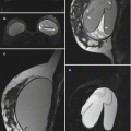
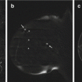
Fig. 5.2
Desmoid tumor local recurrence. (a, b) Sagittal T1-weighted fat-saturated non-contrast image and corresponding T2-weighted fat-saturated image demonstrate an oval, circumscribed T1- and T2-hyperintense mass (arrows) in the lower breast. (c, d) Sagittal post-contrast subtraction image and corresponding CAD enhancement kinetic analysis demonstrate a circumscribed, homogeneously enhancing mass (arrows) with progressive enhancement kinetics
5.3 History
44-year-old with history of pheochromocytoma status post resection 4 years ago, with recent surveillance nuclear medicine scan showing uptake in the left breast. Breast MRI was performed for further evaluation (Figs. 5.3 and 5.4).

Fig. 5.3
Sagittal MR images of the left breast. (a) T1-weighted fat-saturated image. (b) T2-weighted fat-saturated image. (c) T1-weighted fat-saturated post-contrast image. (d) Corresponding CAD color overlay
5.4 Pheochromocytoma
Teaching Points
Pheochromocytomas are an uncommon tumor of the adrenal gland. The tumors generally follow a 10 % rule:
~10 % are extra-adrenal, as illustrated in this case
~10 % are bilateral
~10 % are malignant
~10 % are found in children
~10 % are familial
~10 % are not associated with hypertension
~10 % contain calcification
On MRI, pheochromocytoma has been described as an intensely enhancing mass having characteristic high signal intensity on T2-weighted imaging, typically heterogeneous as illustrated in this case. This “light bulb bright” signal on T2-weighted imaging is neither specific nor sensitive for pheochromocytoma. A characteristic salt-and-pepper pattern has also been described.
Image Findings

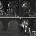
Fig. 5.4
Extra-adrenal pheochromocytoma. Sagittal T1-weighted fat-saturated pre-contrast image (a) and corresponding T2-weighted fat-saturated image (b) of the left breast demonstrate a T1-isointense and heterogeneously T2-hyperintense subareolar mass (arrows). A corresponding T1-weighted fat-saturated post-contrast image (c) demonstrates an intensely homogeneously enhancing 2.9-cm irregular mass (arrow) in the subareolar region, with central washout pattern on CAD kinetics analysis (d)
5.5 History
Screening breast MRI (Figs. 5.5, 5.6, 5.7, and 5.8).

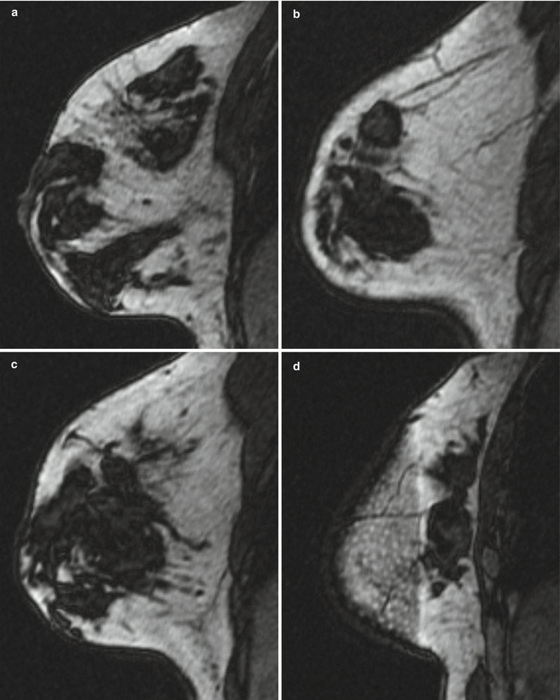

Fig. 5.5
Mammographic views of both breasts. (a) Left craniocaudal (CC) view. (b) Left mediolateral oblique (MLO) view. (c) Right CC view. (d) Right MLO view
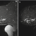
Fig. 5.6
Sagittal MRI images of both breasts. (a–d) T1-weighted images. (e) T2-weighted fat-saturated image. (f, g) T1-weighted fat-saturated pre-contrast and post-contrast images. (h) Image from g with CAD color overlay
5.6 Amyloidosis
Teaching Points
Primary breast amyloidosis is rare, and breast involvement is usually part of the systemic disease. Similar to amyloid deposits elsewhere in the body, calcifications can develop in the breast. Mammographic features of breast amyloidosis include irregular or circumscribed masses with coarse, dystrophic calcifications (Fig. 5.5). On MRI examination, lesions generally demonstrate decreased T1 signal intensity with associated increased T2 signal corresponding with edema and inflammation. Delayed progressive enhancement pattern is usually noted on the post-contrast images. Core biopsy is recommended in most cases of indeterminate findings to confidently exclude co-existent malignancy.
Image Findings

Fig. 5.7
Amyloidosis with associated dense calcifications on mammography, limiting assessment. Left CC (a) and MLO (b) images and right CC (c) and MLO (d) images demonstrate bilateral dense calcifications


Fig. 5.8
Amyloidosis on breast MRI. (a–d) Selected sagittal T1-weighted sequences demonstrate large, conglomerate hypointense areas (arrows) corresponding to areas of dense calcification on mammography. (e) A sagittal T2-weighted fat-saturated image demonstrates corresponding hypointense areas (arrows) with associated peripheral T2 hyperintensity. (f, g) Sagittal T1-weighted fat-saturated pre-contrast and post-contrast images demonstrate mild T1 hyperintensity (arrows) and heterogeneous rim enhancement (arrows) corresponding to the T2-hyperintense regions. No focal suspicious enhancement was identified. (h) CAD enhancement kinetics analysis (arrows) demonstrates a predominantly progressive pattern
5.7 History
Extent of disease evaluation (Figs. 5.9 and 5.10).


Fig. 5.9


Sagittal MR images of the right breast. (a, b) T1-weighted fat-saturated images. (c) T2-weighted fat-saturated image. (d) T1-weighted fat-saturated post-contrast image. (e) Corresponding CAD color overlay image
Related posts:

Stay updated, free articles. Join our Telegram channel
Full access? Get Clinical Tree
