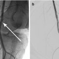
Fig. 33.1
Radiograph of a gasless abdomen and elevated right hemidiaphragm (black arrow)
Ultrasound
Ultrasound cannot rule intra-abdominal hypertension out. However, a useful indicator of increased intraabdominal pressure is elevated Resistive Index, a measure of the resistance to blood flow through arteries. A linear resistive index response to increasing pressure has been described in the renal and hepatic arteries (Fig. 33.2). Evaluation of the morphology and the flow through the inferior vena cava (IVC) and the portal vein may allow for indirect estimation of intraabdominal pressure as well as intravascular volume status and responsiveness to fluid resuscitation. Ultrasound may be very useful for detection of intraperitoneal free fluid, especially large volume ascites, which can be managed at the bedside with directed catheter-based management. Although the reported false negative rate of intra-abdominal free fluid detection is more than 15 % this is more pertaining to small volumes of fluid. Bowel gas and obesity make US studies difficult.
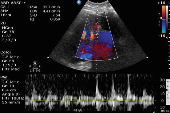
Fig. 33.2


This Color Doppler study of a 48-year-old man who underwent orthotopic liver transplantation shows decreased diastolic flow and high resistive index in the main hepatic artery. Further follow up demonstrated rising intraabdominal pressure secondary to hemoperitoneum
Related posts:


Stay updated, free articles. Join our Telegram channel
Full access? Get Clinical Tree
