INTRACONAL ORBIT: ORBITAL PSEUDOTUMOR AND ACUTE AND CHRONIC INFECTIONS AND NONINFECTIOUS INFLAMMATORY CONDITIONS
KEY POINTS
- The imaging findings in inflammatory conditions involving the muscle cone and central surgical space of the orbit are often nonspecific, but imaging can identify findings based on patterns of involvement that aid in the differential diagnosis of causative pathology in many patients.
- When the muscle cone or central surgical space is involved in infection, there is often some unusual precipitating feature present either clinically or based on inherent patient resistance to infection.
- Imaging can identify causative conditions for infections that can lead to improved outcomes if treated promptly.
- Imaging can identify complications such as detachments, retained foreign bodies, and compressive factors that may lead to improved outcomes if treated promptly.
The noninfectious inflammatory conditions of orbital pseudotumor and the myopathy associated with dysthyroid ophthalmopathy are by far the two most common inflammatory conditions that involve the muscle cone and related central surgical space of the orbit. The effects of dysthyroid ophthalmopathy on the orbit are discussed in Chapter 58 because they are so unique. The pathophysiology of those autoimmune processes and their relationship to other organ systems is discussed in Chapter 20.
Other noninfectious inflammatory diseases may also be local or related to systemic disease. The intraconal compartment is most often secondarily involved due to pathology involving the eye (Chapter 49) and optic nerve/sheath complex (Chapter 56).
Infections may also be local or related to systemic disease. Most of these conditions are due to secondary spread from conditions of eye (Chapter 50), optic nerve and sheath (Chapter 56), and extraconal (Chapter 62) and preseptal compartments (Chapter 70) or from sinonasal disease such as pyogenic sinusitis, rhinocerebral mucormycosis, or other fungal infection (Chapters 84–86). Intraconal infections are also caused by penetrating trauma and surgery.
ANATOMIC AND DEVELOPMENTAL CONSIDERATIONS
Applied Anatomy
The relevant anatomy of the orbit, eye, and optic nerve/sheath complex is discussed in detail in Chapter 44.
IMAGING APPROACH
Techniques and Relevant Aspects
The orbit, eye, and optic nerve and sheath are studied with ultrasound (US), computed tomography (CT), and magnetic resonance (MR) techniques described in detail in Chapters 44 and 45. Specific CT protocols by indications are detailed in Appendix A. Specific MR protocols by indications are outlined in Appendix B. Almost all studies to investigate intraconal pathology are done with contrast.
Pros and Cons
The use of US is for the most part restricted to the identification of a swollen optic disc and possible intraocular complications of disease primarily within the muscle cone. US may show findings immediately adjacent to the nerve–globe junction and around the distal sheath that marginally contribute to the differential diagnosis (Fig. 57.1).
Disease of and within the muscle cone is studied primarily with CT and magnetic resonance imaging (MRI). MRI is far more definitive in its rendering of the optic nerve and ocular effects of such disease and providing objective evidence of secondary optic nerve compromise. MRI is also far more sensitive than CT to a possibly related meningeal pathology. It detects intracranial involvement with these inflammatory processes and their intracranial complications much better than CT. MRI can also screen for related vascular complications and perivascular spread, especially in the cases of fungal disease.

FIGURE 57.1. A: Ocular ultrasound (US) of a patient with posterior uveitis shown as marked thickening of the choroidal layer with fluid in between the choroid and sclera (arrows). There is associated fluid or edema in the Tenon space (arrowheads). B: A patient with posterior scleritis shown as marked localized thickening of the sclera at the posterior pole (arrows). There is fluid in the Tenon space (arrowheads) and along the optic nerve producing the classic US “T” sign.
CT is typically used in very acute situations when an aggressive infectious process, perhaps with a sinonasal origin, or an emergent evaluation of possible associated tension orbit is required. It is also a first choice in patients too sick or for some other reason unable to complete a very high quality, free of motion artifact, MRI examination. CT is a reasonable secondary screening tool for identifying intracranial extension of disease and intracranial complications. Computed tomographic angiography is a good and perhaps preferred screening tool to search for vascular complications such as infectious aneurysms and cavernous sinus thrombosis.
SPECIFIC DISEASE/CONDITION
Orbital Pseudotumor/Idiopathic Orbital Inflammatory Syndrome, and Noninfectious Inflammatory Disease
Etiology
Idiopathic orbital inflammatory syndrome (IOIS) is an acquired immune-based process that lacks systemic involvement.
Other noninfectious inflammatory diseases may also be local or related to systemic disease. The intraconal compartment is most often secondarily involved due to pathology involving the eye (Chapter 49) and optic nerve/sheath complex (Chapter 56). The granulomatoses and histiocytoses are infiltrating orbital processes that may present and appear identical to IOIS or lymphoma. These diseases are discussed in general in chapters on sarcoidosis (Chapter 18), Wegener granulomatosis (Chapter 17), and Langerhans histiocytosis (Chapter 19).
The features of autoimmune processes that may involve the muscle cone and their relationship to other organ systems are discussed in Chapters 20 and 58.
Prevalence and Epidemiology
IOIS is a sporadic disease with no known predisposing factors.
The prevalence and etiology of the granulomatoses, histiocytoses, and autoimmune disease that affect the muscle cone are discussed in the chapters on those entities as noted in the previous section.
Clinical Presentation
Pseudotumor almost always presents as a unilateral swollen and painful eye. There may also be visual loss and ophthalmoplegia. It is occasionally bilateral. The other noninfectious inflammatory diseases are more likely to be bilateral than pseudotumor and the degree of pain and swelling less. Still, the most common cause of unilateral proptosis remains Graves disease.
Pathophysiology and Patterns of Disease
The basic pathology and spread patterns of pseudotumor are discussed in Chapter 18. It is most common in the orbits but occurs elsewhere.
Although IOIS has several morphologic forms, its pathologic basis remains the same; only its distribution varies. These include (a) episcleral or anterior orbital (Figs. 18.15 and 57.2), (b) myositic (Figs. 18.16 and 57.3), (c) perineuritic (Figs. 18.13 and 57.4), (d) diffuse (Figs. 57.5 and 57.6), and dacroadenitic (Fig. 57.3). The actual distribution of the disease frequently spills over from one of these predominant sites of involvement to other areas. The disease most commonly involves the muscle cone and spreads both intraconally and extraconally. Extraocular muscle involvement usually includes the muscle belly, tendon, and sclera adjacent to the tendinous insertion (Fig. 57.3B). Fulminate neoplasms such as leukemia may resemble IOIS both clinically by their rapid onset and imaging pattern. These features help differentiate IOIS from Graves ophthalmopathy or metastases, which typically enlarge only the muscle belly. IOIS can have almost isolated involvement of the Tenon space and be difficult to differentiate from a posterior periscleritis or sclerotenonitis of some other etiology (Chapters 50 and 55).

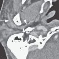
FIGURE 57.2. Contrast-enhanced computed tomography (CECT) of a patient presenting with bilateral orbital swelling, proptosis, and pain. Clinically, the patient was worse on the right than the left. CECT image showing extensive periscleritic swelling (arrows) and findings consistent with vascular congestion (arrowheads). This was a biopsy-proven pseudotumor that responded to steroid therapy.

FIGURE 57.3. Two patients with myositic idiopathic orbital inflammatory syndrome (IOIS). A–C: Patient 1. A patient with mainly myositic pseudotumor. In (A), coronal contrast-enhanced computed tomography reformation shows swelling of the inferior rectus muscle (arrow) and edema within the surrounding intraconal fat (arrowhead). In (B), sagittal reformation shows that the inferior rectus muscle thickening included its tendinous origin and insertion (arrows) compared to normal superior rectus muscle attachments (arrowhead). In (C), the patient also had some minimal periscleritic swelling (arrowheads) as well as inflammation of the lacrimal gland. D: Patient 2. Myositic pseudotumor essentially confined to the lateral rectus muscle. (NOTE: While IOIS may occasionally appear to be isolated to a muscle or one structure in the orbit, most frequently there are other areas of lesser involvement.)
The process is unilateral in at least 85% of cases.1–3 In bilateral cases, it is often asymmetric (Figs. 18.15 and 57.2). Bilateral disease raises the likelihood of an alternative diagnosis of systemic disease such as Graves disease, lymphoma, or sarcoidosis.
Spread to the orbital apex and beyond to the cavernous sinus is possible. Isolated cavernous sinus disease has been called Tolosa-Hunt syndrome (Figs. 18.17 and 57.4–57.5); it is probably reasonable to lump that steroid-responsive granulomatous inflammation of the cavernous sinus into the generalized category of IOIS/pseudotumor.4 Continued spread to the middle and posterior cranial fossa is possible.
Brown superior oblique tendon syndrome is an adherence syndrome, which may be associated with IOIS or other inflammatory diseases or may be congenital. Imaging may show thickening of the tendon and trochlear region with edema in the surrounding fat.
Langerhans cell histiocytoses may involve the orbit in children, usually as a bony lesion with secondary extraconal or transcompartmental orbital extension. The disease may permeate the orbital soft tissues. In the chronic or acute disseminated variants, spread within the optic sheath may be due to seeding from the subarachnoid space of the suprasellar cistern.

FIGURE 57.4. A patient with proven idiopathic orbital inflammatory syndrome of the orbit. This was predominantly of the perineuritic variety. A: Contrast-enhanced T1-weighted fat-suppressed coronal image shows enhancement predominantly around the optic nerve sheath (arrows). B: Swelling extended to the superior orbital fissure (arrows), illustrating the relationship of orbital pseudotumor Tolosa-Hunt syndrome. C: T2-weighted fat-suppressed axial image showing the intraconal swelling mainly around the optic nerve (arrows). There is another area at the orbital apex where the signal is less intense (arrowhead), indicating a more fibrous nature of the disease at the superior orbital fissure.
Related posts:
 Preseptal Compartment: Acute and Chronic Infections and Noninfectious Inflammatory Conditions
 Hypopharynx: Developmental Abnormalities
 Tumors of the Submandibular Gland and Space and Tumorlike Conditions
 Masticator Space, Buccal Space, and Infratemporal Fossa Infections and Other Inflammatory Conditions
 Oral Cavity and Floor of the Mouth: Malignant Tumors
 Benign and Malignant Epithelial Tumors: Lesions of Cutaneous (Skin) Origin
Stay updated, free articles. Join our Telegram channel

Full access? Get Clinical Tree


