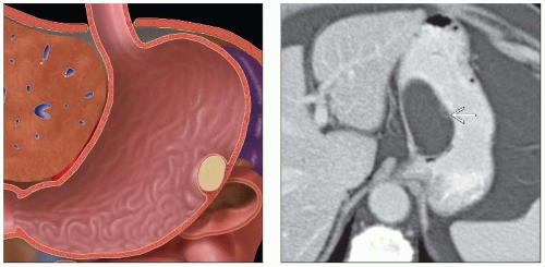
Intramural Benign Gastric Tumors
Michael P. Federle, MD, FACR
Key Facts
Terminology
Benign mass composed of 1 or more tissue elements of gastric wall
50% of all benign tumors are intramural
Remainder are polyps
GIST > lipoma, leiomyoblastoma, lymphangioma, neural tumors
Imaging
UGI series: Intact mucosa, obtuse or right angles with wall
GIST: Often large with central necrosis and ulceration of overlying mucosa on CT
Central area of low attenuation (hemorrhage, necrosis, or cystic formation)
Most GIST > 2 cm have necrosis ± cavitation
Lipoma: Most common in antrum
May prolapse through pylorus into duodenum
Well-circumscribed areas of uniform fatty density = definitive diagnosis
Top Differential Diagnoses
Gastric carcinoma
Gastric metastases and lymphoma
Ectopic pancreatic tissue
Pancreatic pseudocyst
Splenosis
Gastric ulcer
Hematoma/seroma
Diagnostic Checklist
Isolated gastric target lesion = GIST
Multiple target lesions = metastases
TERMINOLOGY
Definitions
Benign mass composed of 1 or more tissue elements of gastric wall
IMAGING
General Features
Best diagnostic clue
Intramural mass with smooth surface and slightly obtuse borders
Other general features
Types of intramural benign gastric tumors
Gastrointestinal stromal tumor (GIST)
Lipoma, leiomyoblastoma, lymphangioma, neural tumors
Radiographic Findings
Upper GI series
Discrete mass, solitary (usually) or multiple
Smooth surface lesion etched in white (double contrast, profile view)
Borders form right angle or slightly obtuse angles with adjacent gastric wall (profile view)
Intraluminal surface of tumor has abrupt, well-defined borders (en face view)
Usually intact overlying mucosa; normal areae gastricae pattern
“Bull’s-eye” or “target” lesions: Central barium-filled crater within mass (ulceration)
± giant, cavitated lesions (GIST)
Pedunculated; may prolapse into duodenum
GIST
Most common; may occur anywhere in GI tract
Several mm to 30 cm
Only 1-2% of GIST are multiple
± extragastric extensions (86%): Gastrohepatic ligament, gastrosplenic ligament, lesser sac
Lipoma, lymphangioma: Tendency to change in size and shape by peristalsis or palpation
Schwannoma and neurofibroma: Multiple lesions with associated abnormalities
CT Findings
GIST
Often large with central necrosis and ulceration of overlying mucosa
Hypo- or hypervascular, well-circumscribed submucosal mass (arterial phase)
Peripheral enhancement (92%)
± homogeneous enhancement (8%)
Central area of low attenuation (hemorrhage, necrosis, or cystic formation)
Stay updated, free articles. Join our Telegram channel

Full access? Get Clinical Tree


