Intramural Pseudodiverticulosis
Michael P. Federle, MD, FACR
Key Facts
Terminology
Rare benign disorder characterized by esophageal diverticulosis-like outpouchings
Caused by dilation of excretory ducts of deep mucous glands
Associated with esophagitis stricture ± motility disorders
Imaging
Barium esophagram more sensitive than endoscopy
Innumerable, tiny, barium-filled outpouchings
In longitudinal rows parallel to long axis of esophagus
Flask-shaped, 1-4 mm
Incomplete filling may erroneously suggest lack of communication with esophageal lumen
Intramural tracks: Bridging between adjacent pseudodiverticula
More likely to occur with diffuse form of intramural pseudodiverticulosis
Stricture: Pseudodiverticula often extend above and below level of stricture
Stricture could be benign or malignant
Top Differential Diagnoses
Esophagitis
Multiple discrete ulcers associated with various types of esophagitis
True ulcers communicate directly with lumen
Ulcers, strictures, and pseudodiverticula can occur together
Diagnostic Checklist
Periodic surveillance of patients with IPD for esophageal carcinoma
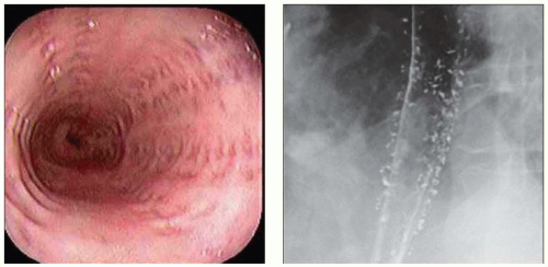 (Left) Endoscopic photograph shows the opening of innumerable pseudodiverticula, many arranged in orderly, longitudinal rows. (Right) Spot film from a barium esophagram demonstrates the classic findings of innumerable tiny collections of barium within the esophageal wall. Decreased primary peristalsis and tertiary contractions were also evident on fluoroscopy. |
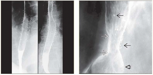 (Left) Two views from a double contrast esophagram reveal innumerable tiny, flask-shaped outpouchings along the entire length of the esophagus, findings pathognomonic of pseudodiverticulosis. (Right) Film from a barium esophagram demonstrates a small hiatal hernia
Get Clinical Tree app for offline access
 and a short peptic stricture and a short peptic stricture
Related posts:Stay updated, free articles. Join our Telegram channel
Full access? Get Clinical Tree


|
