Fig. 11.1
Incidence of dystocia reported in the literature came from 36% to 39%; the graph shows the incidence, in the series. The intrapartum sonography (a) does not currently replace visiting midwife (b) but it is integrated in the obstetrical diagnostic, which is a mental synthesis (c) of obstetric palpation and of the ultrasound images that produce the fetal image in labor by Gifford et al. [2]
However, it shows that on average the two types of delivery amounted to around 35.4% (Fig. 11.2).
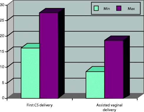
Fig. 11.2
Incidence of caesarean section in the USA (first CS delivery) and operative delivery (assisted vaginal delivery in the USA): the columns indicate the minimum and maximum of the two types of delivery (Hanley et al. [20])
The different types of delivery have undergone an interesting evolution over the past twenty years.
The inexorable increase of caesarean section is associated with a considerable reduction in the use of the obstetric vacuum and especially of the forceps (Fig. 11.3).
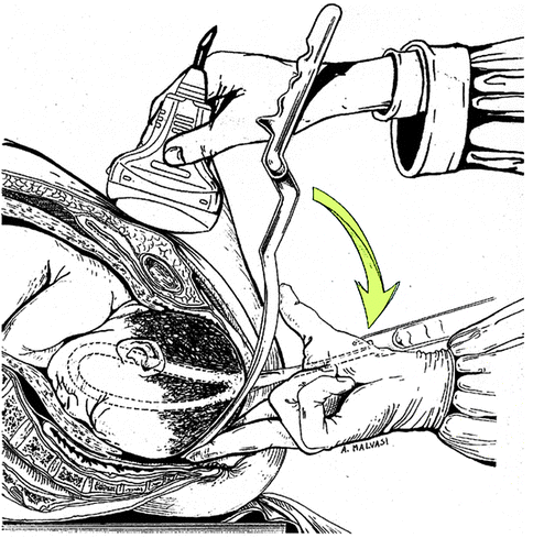
Fig. 11.3
Application of forceps direct and symmetric in occipital-anterior under ultrasound guidance
One reason for the increase in the caesarean section was the patient’s request, even in the absence of traditional indications. The reasons for these requests are the fear of perineum damages (urinary and fecal incontinence, genital prolapse), but also the reduction of fetal risks due to vaginal delivery.
Regarding the use of forceps and the obstetric vacuum, while the effectiveness of these instruments is still recognized, but considering the severity of maternal and fetal injuries resulting from their use, many practitioners prefer to limit their use without very clear indications.
The clinical risk management in relation to fetal distress due to hypoxia or asphyxia during birth remains a central point as a major cause of coroner’s action and increased costs for operators and health facilities.
The incidence of encephalopathy and cerebral palsy remains unchanged in industrialized countries than in developing countries despite the obstetricians’ progress, such as continuous cardiotocographic monitoring [21].
The ACOG (American College of Obstetricians and Gynaecologists) reports that the percentage of cerebral palsy associated with intrapartum events is around 10%. Acute fetal asphyxia is less associated with adverse neonatal outcomes compared to prolonged intrauterine hypoxia. The current opinion is that severe perinatal asphyxia has not poor prognosis because an adequate neonatal resuscitation can avoid most of the brain damage in babies and that most of the survived infants does not have adverse neurological outcomes.
In contrast, chronic asphyxia has greater incidence of permanent damages [22].
In order to quickly resolve the dystocial labors at risk of acute asphyxia is therefore crucial to the proper use of forceps or vacuum cup, especially in cases of dystocia.
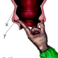
The placement of the cup in case of application of the vacuum or the application of forceps, in cases of asynclitism are two situations in which the operator finds good help with intrapartum sonography (Fig. 11.4).
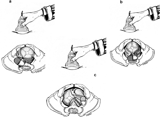
Fig. 11.4
Application of forceps under ultrasound guidance: (a) straight, and symmetrical in occipitoposterior right position; (b), straight, and symmetrical occipitoposterior position of the median; (c) direct, and symmetrical occipitotransverse left position
The application of the obstetric vacuum is closely related to the correct determination of the position of the fetal head because otherwise it causes a deflection of the fetal head with consequent probable detachment of the cup itself and increase in neonatal morbidity.
The application of the obstetric vacuum on deflected occiput increases by 4–5 times the probability of failure, while its application on posterior or lateral occiput increases two times the chances of failure [23].
If the fetal head is deflected at the time of the application of the obstetric vacuum, the probability of a low Apgar score (less than 6) increases by 3.2 times and the probability of severe trauma of the scalp of 5.2 times and increases 12 times the probability of admission to neonatal intensive care [24, 25].
Since the lateral and posterior positions of the most difficult to diagnose with an obstetric examination, and as reported in the literature these situations have a higher frequency with epidural analgesia, the use of ultrasound gets more important with operative delivery during labor analgesia [26].
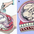
In case of vaginal operating delivery, the intrapartum sonography can therefore allow a more precise application of the forceps and of the vacuum, in particular for the latest disposable cups (Kiwi), which, if not positioned properly, can easily detach especially in case of misapplication over caput succedaneum (Fig. 11.5).
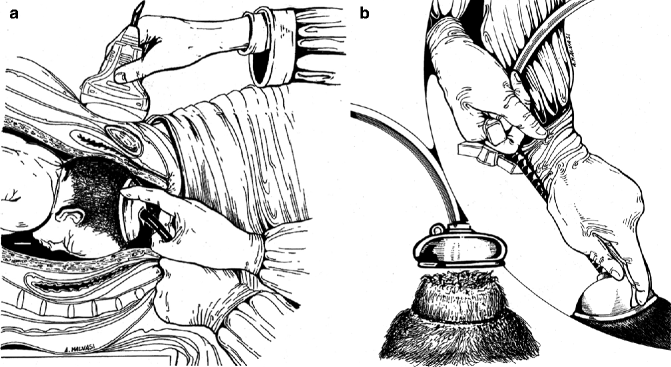
Fig. 11.5
Application of obstetric vacuum with ultrasound guide (a). Application of the obstetric vacuum on caput succedaneum and subsequent depression of the cup on the fetal head (b). If possible, the obstetric vacuum should be applied far away from the caput succedaneum, under ultrasound guidance
The intrapartum sonography is particularly important in cases of dystocia delivery, as it provides an image that guides the application of the branches of the forceps.
In particular, in anterior or posterior asynclitism, if you decide to apply the forceps, intrapartum transabdominal ultrasound can guide the direct and symmetrical application on the parietal bones, avoiding improper application of the branches on the face. The trans-labial ultrasound instead allows assessing the level of the presenting part, letting you to decide if there are the conditions for the application of the forceps or not.
In particular, in anterior or posterior asynclitism, if you decide to apply the forceps, intrapartum transabdominal ultrasound, can guide the direct and symmetrical application on the parietal bones, avoiding improper application of the branches on the face. The trans-labial ultrasound instead allows to assess the level of the presenting part, letting you to decide if there are the conditions for the application of the forceps or not.
Similarly, the application of the cup can be performed under ultrasound guidance highlighting the position of the fetal head and the localization of the caput succedaneum, avoiding the positioning of the cup on the tumors and prevent failures, as reported in different studies by Molina and Nicolaides [26], Sentilhes et al. [27], and Lau et al. [28].
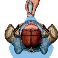
Even in the case of caesarean sections, intrapartum sonography is also useful to consider other important intra- and postoperative data. For example, in occiput-posterior presentations, with ruptured membranes and with fixed head in the pelvic cavity, we should consider the position of the fetal head in order to avoid fetal injuries by cuts on the face. Nevertheless, in case of greatly flexed head, a pre\operative ultrasound evaluation is useful in order to prevent incongruous maneuvers during the extraction of the head (Fig. 11.6).
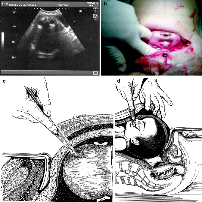
Fig. 11.6
Caesarean section with fetal median occiput-posterior position. Image diagnosed with transabdominal intrapartum ultrasound (a) and intraoperative corresponding after the hysterotomy (b). In occiput-posterior position and ruptured membranes iatrogenic injuries over the fetal head are possible when the uterine incision is not made with great caution: cold knife scalpel lesion on the scalp (c), scarring on the face of the fetus (d)
The intrapartum use of ultrasound is very important in early diagnosis of dystocia, which avoids unnecessary prolonged labors that often end with emergency caesarean sections (with a higher percentage of intra- and postoperative complications).
In particular, significant intraoperative bleeding complications may be due to an excessive distension of the lower uterine segment [29–36]. Postoperative complications may also occur after an inadequate hemostasis, but also as a consequence of hypotension caused by spinal anesthesia (Fig. 11.7) which is followed by a normalization of blood pressure that could generate a bleeding site if eschars fall from the vessels treated with electrocoagulation. This phenomenon could mainly happen in hypertensive or preeclamptic patients [37, 38].
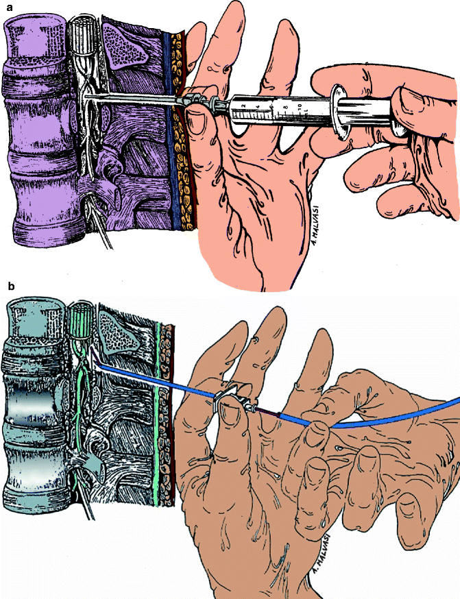
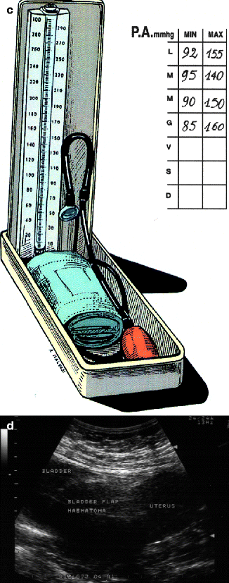
Fig. 11.7
The spinal and epidural anesthesia (a) or combined spinal epidural – CSE (b), commonly used for caesarean section, cause hypotension; therefore, inaccurate hemostasis may develop a later bleeding when the pressure returns to normal (c), causing possible complications of uterine suture as a “bladder flap hematoma” (d). Ultrasound image of bladder flap haematoma (from Tinelli A et al, Gynecol Surg (2007) 4: 53–56. DOI 10.1007/s10397-006-0212-2.)
Recent studies have shown that prolonged dystocial labor is caused not only by mechanical fetus-pelvic causes but also by dysfunctions of the uterine contractility that are associated with dynamic dystocia. In particular, the dystocial labor is also associated with an alteration of neurotransmitters that modulate the activity of the upper and lower uterine segment.
Also, excessive distension of the lower uterine segment by the presented part of the fetus changes the neurotransmitters creating a vicious cycle that increases the degree of dystocia, causes pain, and increases blood circulation. This provokes a more vascularized uterine segment, with possible intraoperative difficulties, and a reduction of neurotransmitters that accentuate the dystocia. In the uterine scar that is created, we have a reduced function of neurotransmitters in a subsequent labor, especially if you program a vaginal birth after caesarean section (VBACS) [39].
Another aspect of bleeding complications in the course of CS, the postpartum and early puerperium are related to the surgical technique used.
In particular, the suturing of the visceral peritoneum in the case of bleeding causes the “bladder flap hematoma” but in case of bleeding without suturing, the bladder fold can cause a hemoperitoneum [29–32] (Fig. 11.8).
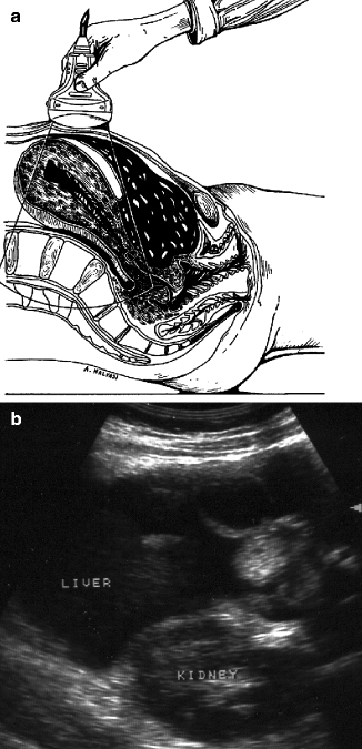
Fig. 11.8
Hemoperitoneum in postpartum after caesarean section without suturing the parietal peritoneum. (a) Drawing representing hemoperitoneum after caesarean section, (b) corresponding US image (Amended by Malvasi et al. [34])
Similarly, in case of bleeding with suture of the parietal peritoneum, a subfascial hematoma can develop (Fig. 11.9), but without the suture of the peritoneum, leaving the Retzius space open, the hemoperitoneum can form [33, 40].
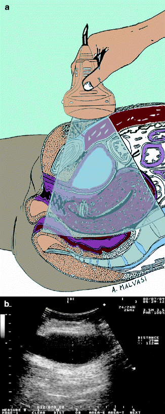
Fig. 11.9
Subfascial hematoma after caesarean section with the parietal peritoneum sutured. (a) Drawing representing subfascial hematoma after caesarean section, (b) corresponding US image (Amended by Malvasi et al. [33])
11.1 Management of Obstetric Risk in Thrombophilic Patients
The use of intrapartum sonography and in particular the trans-labial reduces the number of caesarian sections in favor of vaginal operative deliveries with the vacuum cup, as reported in a study of Dukelman et al. [41].
The intrapartum sonography always improves the diagnosis, especially of the position of the fetal head, but also of its descent and rotation. It is also particularly useful in labors of pregnant women with systemic diseases, such as hypertension and diabetes.
Another interesting aspect of intrapartum ultrasonography is in the management of labor in patients with thrombophilia, where a spontaneous delivery is indicated (that has less risk than operative delivery).
The Enquiry Maternal Death 2005–2008 [42] has demonstrated over the previous three years that a screening action and therapy in thrombophilic patients has reduced maternal deaths at delivery and puerperium.
In fact, thrombosis is a potentially serious complication that can occur during pregnancy. It can manifest against the mother, in the form of venous thromboembolic disease, or it can develop an obstetrical complication (placental thrombosis).
The relative risk of thromboembolism during pregnancy has increased about 10 times. This number must be taken with extreme caution because this is compared with a very low absolute incidence data of venous thromboembolism in nonpregnant women of similar age.
In fact, if a case of thromboembolism occurs every 10,000 women in this age group, during pregnancy the incidence rises to about one in 1,000 pregnant women. So, in absolute terms, we cannot speak about a common complication [43] (Fig. 11.10).
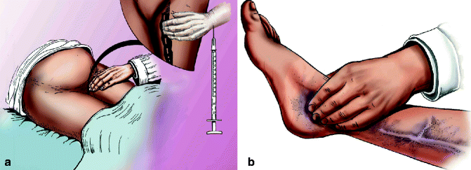
Fig. 11.10
Femoral vein thrombosis in pregnancy and trophic changes of the leg and foot. (a) Image representing Scarpa’s triangle, which runs the femoral vein. (b) Image representing the altered trophism of the lower limb
However, we have to keep in mind that pulmonary embolism remains the most important and most frequent causes of maternal death.
The clinical presentation of VTE during pregnancy is unusual, especially for what concerns the deep vein thrombosis.
The location of deep venous thrombosis in a high percentage of cases, about 85%, occurs against the left lower limb. Furthermore, it is much more frequent that the thrombosis is localized to the femoral iliac district affecting distal veins, then the veins of the leg in only about 10% of cases [44].
This is attributable to the effect of stasis related to mechanical compression of the gravid uterus.
Semiotics of deep vein thrombosis in pregnancy may be unusual.
In fact, it can manifest as abdominal pain in the lower quadrants and fever.
In a high percentage of cases, episodes of venous thromboembolism in pregnancy are associated with the presence of risk factors.
These risk factors have been identified with age of women during pregnancy (over 35), the need to perform a caesarean section, obesity, family history, and even more with the personal history of venous thromboembolism. Also, venous thromboembolism seems to be more frequent in multiparous women, compared with primiparous.
Other risk factors include ovarian hyperstimulation in cases of medically assisted procreation (MAP), prolonged immobility during pregnancy, and the presence of varicose veins.
For inherited thrombophilia, we mean a high risk of developing venous and/or arterial thrombosis, mostly at a young age (<50 years), not easily linked with obvious risk factors, with a tendency to recur. Thrombophilia is also a hypercoagulable prothrombotic condition. The thrombotic episodes are more frequent in venous districts than arterial.
Related posts:
 General Intrapartum Sonography Setup and Use in Labor
 New Technologies for Monitoring Labor Progress
 Use of Cervical Length in Labor and Delivery
 Clinical Evaluation of Labor and Intrapartum Sonography
 Occiput Posterior Position and Intrapartum Sonography
 Intrapartum Sonography and Labor Progression
Stay updated, free articles. Join our Telegram channel

Full access? Get Clinical Tree


