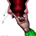
Fig. 9.1
Correct palpation of head station in an obstetrical phantom, expressed in centimeters above or below the level of the ischial spines, as assessed by 32 residents and 25 consultant obstetricians (Data from [11])
9.2 Translabial Ultrasound
Different descriptive terms have been used for the insonation of the birth canal from below the pubic symphysis, for which the transducer is placed between the pubic bones and the anus: translabial, perineal, and transperineal. “Translabial ultrasound” describes best how the transducer is actually applied: A (curved array) transducer that is typically used for a transabdominal mid- and third-trimester exam is placed on the labia, sagittally in a median plane. In the top of the image and close to the entrance, a longitudinal section of the pubic symphysis is displayed and the perineum (the anatomical region between the posterior commissure and the anus) is on the right (Fig. 9.2a, b).
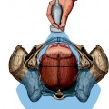
Fig. 9.2
Comparison of abdominal and translabial ultrasound images. (a1) Median transabdominal longitudinal section cranial to the pubic symphysis, showing a dorsoanterior, occipito-anterior cephalic lie. (a2) Transabdominal ultrasound evaluation technique shows the fetal head position in longitudinal scan on the first stage of labor, of a fetus in cephalic presentation, and occipito-transverse position. (b1) Intrapartum translabial ultrasound ITU (the pubic symphysis is not visible). The transducer is placed median on the labia in such a way that the pubic symphysis is cut in the midline and lies horizontally on the top of the image. The fetal head is easily recognized, and the direction of its longest axis with regard to the axis of the curved birth canal (the so-called head direction) is apparent. The dorsal part of the birth canal (soft tissue and sacral bones) cannot be delineated well (Images with permission from [12]). (b2) “Head down”: ultrasound translabial schematic sagittal scan representing the direction of the fetal head down. We distinguish the pubic bone and the outline of the fetal head with evident tumor from birth
The term “perineal” or “transperineal” insonation is ambiguous because in English textbooks, different definitions for “perineum” exist one of which also includes the vulva. Hence, for clarity the term, “intrapartum translabial ultrasound” or “ITU” should be used.
9.3 Obtaining ITU Images
A curved array transducer such as typically used for mid- and third-trimester transabdominal ultrasound is used for ITU, but placed median on the labia yields a “panoramic” view of the birth canal. In this easy to obtain plane, the bony structures that define the head station cannot be seen: the ischial spinae define the narrowest plane of the bony birth canal and indicate head station ±0 cm. However, there is a fixed anatomical relationship between the pubic symphysis and the ischial plane: In a median section, line perpendicular to the inferior border of the pubic symphysis is 3 cm above the parallel plane through the ischial spines on both sides [6, 13].
Using this reference system, the head station determined objectively by ultrasound can be directly compared and related to the “classical” coordinates of vaginal obstetrical palpation, expressing the station of the presenting part in centimeters above or below mid-pelvis.
From a clinical perspective, the dynamic aspect of ITU are important, same as during a palpation when the clinician assesses the head station at rest and at the height of pushing during a naturally occurring contraction. These dynamics can be observed also using ITU and can even be quantified (Fig. 9.3). From a practical point of view, the image can be optimized in the absence of a contraction and while the woman is resting. When the contraction peaks, the next image should be taken. Then, the woman can be told to push, and another image is taken. Finally, the parameters described should be analyzed for these three situations to enable recognition of dynamic changes (Fig. 9.3).
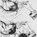
Fig. 9.3
Descent of the fetal head as seen on infrapubic intrapartum ultrasound. The long sides of the yellow boxes indicate the infrapubic line (on the left) and the level of the ischial spines (on the right). The axis of the upper short side of the box is coincident with the long exis of the pubic symphysis. (a) resting, without contraction, (b) at the height of a contraction, (c) with pushing during contraction, and (d) after a further contraction and immediately prior to a successful vacuum extraction (modified after 12)
9.4 Parameters of Intrapartum Translabial Ultrasound
The quantitative parameters of ITU (Table 9.1) are all based on an exact median section of the pubic symphysis (Fig. 9.4). The leading landmark is the lower end of the symphysis or, more specifically, a perpendicular line extending dorsally; this line can easily be visualized in all patients; it is called the “infrapubic line.”
Table 9.1
Parameters used to describe birth progress as seen on intrapartum translabial ultrasound (ITU)
ITU parameter | Comments |
|---|---|
Head station | Corresponds to the well-established palpated head station, is also expressed in centimeters above or below mid-pelvis as defined by the ischial spines |
Head direction | A new parameter that describes the physiological direction of the fetal head as it descends through the birth canal |
“Angle of descent” (also called “angle of progression”) | Easy to learn and assess, correlates with ITU head station in a linear fashion |
Descent during a contraction (±augmentation by pushing) | Includes assessment of the dynamics of the birth “powers” |
Fig. 9.4
Possible objective parameters of intrapartum translabial ultrasound (ITU). All quantitative parameters require an exact median section of the pubic symphysis. The main landmark is the caudal end of the symphysis or rather a perpendicular line extending dorsally, the so-called infrapubic line. This infrapubic lines run 3 cm cranial to a parallel plane that traverses the ischial spines, which in turn define mid-pelvis (station ±0 cm). In addition, reference to the long axis of the symphysis enables angle measurements, for example, that of the head direction (direction of the longest visible axis of the fetal head in the birth canal as seen in this insonation) as well as placing a tangent on the deepest bony part of the fetal head; together with the long axis of the pubic symphysis, this tangent defines the “angle of descent” or “angle of progression.” Note: During a normal birth, almost always a scalp edema (caput succedaneum) becomes visible (in the image indicated by “*”) which must not be confused with the skull
The ischial spines are the anatomical structures whose level along the birth canal defines mid-pelvis (station ±0 cm); however, they cannot be seen on ITU. CT studies have shown that the infrapubic lines runs 3 cm cranial to a parallel plane that traverses the ischial spines [6].
Because, practically, there is a fixed anatomical relationship between the ischial spines and the pubic bone, the pubic symphysis can be used to calculate the level of the ischial spines: Parallel planes through the infrapubic line and the ischial spines are 3 cm apart, and measurements relating to the infrapubic line – which is easily seen on ITU – can be converted into the established obstetrical system of centimeters above or below the ischial spines.
In addition, reference to the long axis of the symphysis enables angle measurements, for example, that of the head direction (direction of the longest visible axis of the fetal head in the birth canal as seen in this insonation) as well as placing a tangent on the deepest bony part of the fetal head; together with the long axis of the pubic symphysis, this tangent defines the “angle of descent” or “angle of progression.”
As the fetal head descends through the birth canal, the angle between the symphysis axis and the longest axis of the fetal head visible on ITU changes direction: While it may be “down” to “horizontal” in the upper part of the birth canal, it eventually “homes” towards an upward direction (>20°) once the head has passed the mid-pelvis. This relatively simple qualitative assessment for the “head-up sign” seems to be important when deciding operative vaginal delivery (see below).
Related posts:
Stay updated, free articles. Join our Telegram channel

Full access? Get Clinical Tree


