Introduction to Breast Elastography
Ultrasound evaluation of the breast was initially used to determine if a lesion was cystic or solid.1,2 With the criteria set forth by Stavros et al,3 ultrasound has since had a more important role in breast lesion characterization. These criteria have been incorporated into the ultrasound Breast Imaging–Reporting and Data System (BI-RADS) lexicon4 and can be used to distinguish between benign and malignant breast lesions.
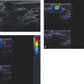
Elastography is a new ultrasound technique that can provide additional information which was previously not available. Elastography or elasticity imaging (EI) is an imaging modality based on tissue stiffness, rather than anatomy. These images show the relative difference in stiffness among tissue. For thousands of years physicians have used palpation for diagnosis of breast cancer,5 realizing that stiffer masses on palpation were more likely malignancies. Ultrasound elastography has the potential to quantify the stiffness of a lesion, which was previously judged only subjectively by physical exam.6–8 Krouskop et al determined that in vivo there is significant elastographic contrast between cancerous and noncancerous breast lesions.9 This suggests that elastography should be an excellent technique for characterizing breast lesions as benign or malignant.
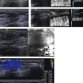
There are two types of elastography: strain elastography (SE) and shear wave elastography (SWE). Strain elastography produces an image based on the displacement of the tissue from a compression/release force applied by an external force (transducer or acoustic radiation force impulse [ARFI]) or a patient source (breathing and/or heartbeat). This allows for a qualitative assessment of the lesion, that is, a relative assessment of the stiffness compared with other tissues in the field of view. The exact stiffness of the lesion is not obtained. SWE applies a special “push pulse,” ARFI, which results in shear wave propagation that can be measured as a velocity. Because the velocity of the shear wave through tissues is dependent on the “stiffness” of the tissue, a quantitative value of the stiffness can be obtained; that is, a measurement of lesion stiffness is obtained and expressed as a numerical value. With the addition of elastography we now have three ultrasound modes (▶ Table 1.1).
| Mode | What is measured: | What is displayed: |
| B-mode | Acoustic impedance | Anatomy |
| Doppler | Motion | Vascular flow |
| Elastography | Mechanical properties | Tissue stiffness |
Related posts:
Stay updated, free articles. Join our Telegram channel

Full access? Get Clinical Tree


