Chapter 1 Introduction to diagnostic ultrasound
AUDIBLE SOUND
Sound is a form of energy which causes a mechanical disturbance in the form of vibration of molecules within a medium. In order to be transmitted, sound requires a medium containing molecules, and therefore cannot travel through a vacuum. The production of sound requires a vibrating object, such as a tuning fork, which when physically struck will vibrate. It will then cause adjacent air molecules to vibrate, and these in turn will cause their neighboring molecules to vibrate. This disturbance will spread through the air as a longitudinal wave. This means that the wave travels from the source of the vibration, parallel to the direction in which the particles vibrate. The phase of the wave when the molecules are pushed together is called compression, and when apart, rarefaction.
ULTRASOUND
Ultrasound is the name given to high-frequency sound waves, which are above the human hearing range. Diagnostic ultrasound travels in a similar way to audible sound. It consists of minute mechanical vibrations (pulses of ultrasound) which are transmitted into the body. As the ultrasound wave propagates (travels) through the body, it causes a local displacement of molecules within the medium. Figure 1.1 shows the changes occurring within a medium as the sound travels through it.

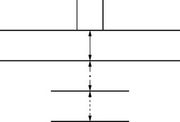
The point at which the tissue type changes (the interface) is where there is a change in acoustic impedance (and a change in speed of travel of the sound), and this will cause part of the pulse to be reflected back in the form of an echo, with the remainder traveling on through the body (see Fig. 1.2). The larger the difference in acoustic impedance between two tissues, the more sound will be reflected back to the transducer and the less sound will carry on traveling through the tissue. These returning echoes are converted into a visual display and used to form a sectional image. This sequence of events is known as the pulse-echo principle.