Introduction
The available imaging arsenal for the detection and management of ischemic heart disease has never been larger. Many new noninvasive imaging techniques have obviated the need for many invasive diagnostic procedures and offer risk stratification of patients with known and newly detected disease. Imaging allows the stratification of patients with preclinical disease, beyond that of clinical risk factors. Finally, in cases of invasive diagnostic workups, new imaging modalities now offer highly accurate quantification and adjunctive information that was previously not possible.
The Ischemic Cascade: A Framework for Categorization
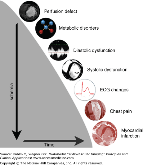
Given the often overwhelming array of imaging modalities and potential findings from each given imaging test, a framework with which to categorize imaging findings is useful. The “ischemic cascade” that follows coronary artery occlusion (Fig. 20–1), as first described by Nesto and Kowalchuk,1 provides such a framework. By establishing the progression from clinically unrecognized parameters to myocardial infarction, the concept of the ischemic cascade allows the physician to recognize and categorize the potential findings from a given imaging study and interpret them along a clinically meaningful spectrum.
Figure 20–1.
Following coronary stenosis or occlusion, a progression of detectable abnormalities of the myocardium begins with decreased myocardial blood flow (perfusion defects) at rest or during stress (exercise or pharmacologically induced). Diastolic myocardial dysfunction then follows, with later systolic dysfunction. Eventually, electrocardiogram (ECG) changes and progression to infarction occur. Various imaging tests can detect or infer these abnormalities.
The progression along the continuum of detectable abnormalities of the myocardium is preceded by detectable precursors affecting the coronary arteries, namely atherosclerotic changes, although simple coronary arterial disease may not lead to ischemic consequences. In the presence of a discrete coronary artery stenosis or generalized arteriopathy significant enough to cause significantly decreased myocardial blood flow at rest or during stress (exercise or pharmacologically induced), perfusion defects can be initially detected. Diastolic myocardial dysfunction follows and then systolic dysfunction. Eventually, electrocardiogram (ECG) changes and infarction can be detected. Various imaging tests can detect or infer the presence of myocardial ischemia or infarct. These findings are summarized in Table 20–1.
| Modality | Echo | CT | MRI | SPECT | PET | SCA |
|---|---|---|---|---|---|---|
| Plaque | − | +++ | + | − | − | ++ |
| Perfusion defect | − | + | +++ | ++ | +++ | − |
| Diastolic dysfunction | +++ | + | +++ | − | − | − |
| Systolic dysfunction | +++ | ++ | +++ | + | + | + |
| Infarction | + | ++ | +++ | ++ | ++ | + |
| Other findings | +++ | +++ | +++ | + | + | + |
Therefore, in this chapter, we discuss each imaging modality and outline the potential findings offered by each modality along the preclinical and clinical sequence of the ischemic cascade. When selecting the appropriate test for a given patient, the multisociety appropriateness criteria can serve as guidelines, allowing evidence-based expert consensus to guide the clinician in evaluating complex imaging decisions in an efficient manner.2-5 In many cases, the complementary information provided by multiple tests is essential to patient management.6 In certain instances, hybrid imaging allows simultaneous acquisition of this information (ie, positron emission tomography [PET]/computed tomography angiography [CTA] or single photon emission computed tomography [SPECT]/computed tomography [CT]).7
Radiography/Fluoroscopy
Chest radiography is a universally available, relatively inexpensive test that allows reliable assessment of cardiac size and of the sequelae of ischemic heart disease such as pulmonary venous hypertension, pulmonary edema from cardiogenic origin, and focal edema from ischemic valve disease (such as mitral regurgitation). Although normal radiographs cannot exclude ischemic heart disease, this easily performed test can reliably detect changes over time in patients with known heart disease.8 X-ray fluoroscopy has historically been used to detect coronary calcification and cardiac size but is no longer used due to the availability of more reliable methods.
Ultrasound
Cardiac ultrasound, or echocardiography, is a widely available, radiation-free modality that is routinely used in the diagnosis and management of all forms of heart disease. Transthoracic echocardiography is the most commonly performed imaging examination in the United States.9 Although limited by operator-dependent techniques, acoustic windows, and body habitus in obese patients, echocardiography plays a key role, offering robust information without the use of iodinated contrast or ionizing radiation. Transesophageal echocardiography, an invasive test, allows more reliable acoustic windows and imaging planes at the expense of time and invasiveness. Echocardiography allows the assessment of ventricular and atria size, global and regional left ventricular function, diastolic function (tissue relaxation/ventricular filling), myocardial wall thickness, and assessment for valvular disease. Doppler ultrasound also allows measurement of flow and calculation of gradients, such as in cases of valve dysfunction.3 New techniques in echocardiography such as deformation (tissue tracking imaging), application of contrast, and three-dimensional imaging can expand the potential role of echocardiography and aid in selected cases.
Stress Echocardiography
Stress echocardiography, performed with dobutamine (pharmacologic stress) and/or low-level exercise, allows the detection of induced wall motion abnormalities, a key finding along the ischemic cascade. This finding alone results in a high diagnostic accuracy for the detection of significant coronary artery disease (CAD).2 Two different meta-analyses have been performed comparing the performance of stress echocardiography to conventional coronary angiography. The first included patients with an intermediate pretest likelihood of CAD (25%-75%), and the angiographic definition of significant coronary heart disease was either ≥50% or ≥70%. Stress echocardiography had a sensitivity and specificity of 76% and 88%, respectively, in six studies of 510 patients.10
The second meta analysis compared exercise echocardiography and exercise SPECT myocardial perfusion imaging with coronary angiography, with extent of stenosis varying between ≥50% and 75%. The two tests had similar sensitivity (85% for echocardiography and 87% for SPECT), but the specificity for stress echocardiography was significantly higher (77% for echocardiography vs 64% for SPECT).11
Computed Tomography
CT of the heart has been performed for decades, but the challenges of cardiac motion and ECG gating have until recently been daunting. Early success with electron-beam CT allowed reliable quantification of calcium scores, which highly correlate with (preclinical) CAD burden.12 However, this expensive technology has not become widely available.
With the advent of multidetector CT (MDCT), routine assessment of the coronary arteries became widely available. MDCT offers reliable calcium scoring. The use of calcium scoring can be used to lower the likelihood of CAD, but numerous recent studies have shown that a significant proportion of patients have only noncalcified plaque, which will be missed by calcium scoring CT.13
Stay updated, free articles. Join our Telegram channel

Full access? Get Clinical Tree


